Iron »
PDB 1tfz-1ubh »
1tqn »
Iron in PDB 1tqn: Crystal Structure of Human Microsomal P450 3A4
Enzymatic activity of Crystal Structure of Human Microsomal P450 3A4
All present enzymatic activity of Crystal Structure of Human Microsomal P450 3A4:
1.14.14.1;
1.14.14.1;
Protein crystallography data
The structure of Crystal Structure of Human Microsomal P450 3A4, PDB code: 1tqn
was solved by
J.K.Yano,
M.R.Wester,
G.A.Schoch,
K.J.Griffin,
C.D.Stout,
E.F.Johnson,
with X-Ray Crystallography technique. A brief refinement statistics is given in the table below:
| Resolution Low / High (Å) | 50.00 / 2.05 |
| Space group | I 2 2 2 |
| Cell size a, b, c (Å), α, β, γ (°) | 77.153, 99.615, 132.684, 90.00, 90.00, 90.00 |
| R / Rfree (%) | 23.8 / 29.3 |
Iron Binding Sites:
The binding sites of Iron atom in the Crystal Structure of Human Microsomal P450 3A4
(pdb code 1tqn). This binding sites where shown within
5.0 Angstroms radius around Iron atom.
In total only one binding site of Iron was determined in the Crystal Structure of Human Microsomal P450 3A4, PDB code: 1tqn:
In total only one binding site of Iron was determined in the Crystal Structure of Human Microsomal P450 3A4, PDB code: 1tqn:
Iron binding site 1 out of 1 in 1tqn
Go back to
Iron binding site 1 out
of 1 in the Crystal Structure of Human Microsomal P450 3A4
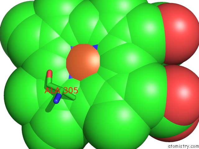
Mono view
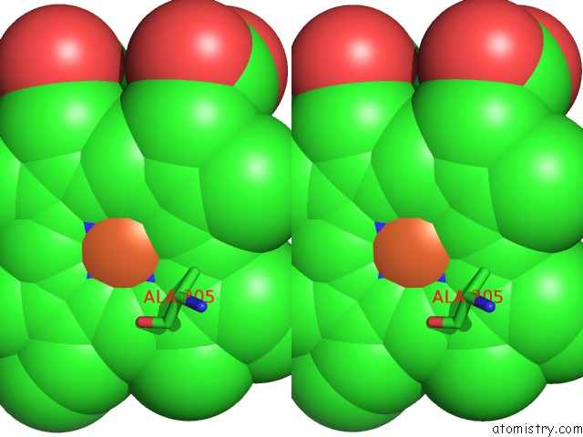
Stereo pair view
Mono view
Stereo pair view
A full contact list of Iron with other atoms in the Fe binding
site number 1 of Crystal Structure of Human Microsomal P450 3A4 within 5.0Å range:
|
Reference:
J.K.Yano,
M.R.Wester,
G.A.Schoch,
K.J.Griffin,
C.D.Stout,
E.F.Johnson.
The Structure of Human Microsomal Cytochrome P450 3A4 Determined By X-Ray Crystallography to 2.05-A Resolution J.Biol.Chem. V. 279 38091 2004.
ISSN: ISSN 0021-9258
PubMed: 15258162
DOI: 10.1074/JBC.C400293200
Page generated: Wed Jul 16 21:04:26 2025
ISSN: ISSN 0021-9258
PubMed: 15258162
DOI: 10.1074/JBC.C400293200
Last articles
Na in 7U68Na in 7U34
Na in 7UCC
Na in 7UC8
Na in 7U59
Na in 7U1R
Na in 7U0O
Na in 7U0M
Na in 7TXS
Na in 7U1C