Iron »
PDB 1ubj-1uvy »
1uof »
Iron in PDB 1uof: Deacetoxycephalosporin C Synthase Complexed with Penicillin G
Enzymatic activity of Deacetoxycephalosporin C Synthase Complexed with Penicillin G
All present enzymatic activity of Deacetoxycephalosporin C Synthase Complexed with Penicillin G:
1.14.20.1;
1.14.20.1;
Protein crystallography data
The structure of Deacetoxycephalosporin C Synthase Complexed with Penicillin G, PDB code: 1uof
was solved by
K.Valegard,
A.C.Terwisscha Van Scheltinga,
A.Dubus,
L.M.Oster,
G.Rhangino,
J.Hajdu,
I.Andersson,
with X-Ray Crystallography technique. A brief refinement statistics is given in the table below:
| Resolution Low / High (Å) | 56.8 / 1.6 |
| Space group | H 3 |
| Cell size a, b, c (Å), α, β, γ (°) | 106.500, 106.500, 72.100, 90.00, 90.00, 120.00 |
| R / Rfree (%) | 19.6 / 23.1 |
Iron Binding Sites:
The binding sites of Iron atom in the Deacetoxycephalosporin C Synthase Complexed with Penicillin G
(pdb code 1uof). This binding sites where shown within
5.0 Angstroms radius around Iron atom.
In total only one binding site of Iron was determined in the Deacetoxycephalosporin C Synthase Complexed with Penicillin G, PDB code: 1uof:
In total only one binding site of Iron was determined in the Deacetoxycephalosporin C Synthase Complexed with Penicillin G, PDB code: 1uof:
Iron binding site 1 out of 1 in 1uof
Go back to
Iron binding site 1 out
of 1 in the Deacetoxycephalosporin C Synthase Complexed with Penicillin G
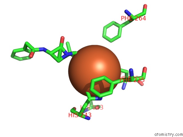
Mono view
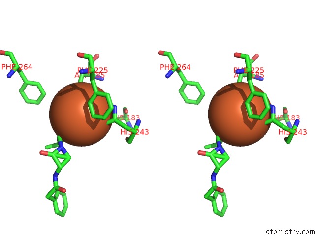
Stereo pair view
Mono view
Stereo pair view
A full contact list of Iron with other atoms in the Fe binding
site number 1 of Deacetoxycephalosporin C Synthase Complexed with Penicillin G within 5.0Å range:
|
Reference:
K.Valegard,
A.C.Terwisscha Van Scheltinga,
A.Dubus,
G.Ranghino,
L.M.Oster,
J.Hajdu,
I.Andersson.
The Structural Basis of Cephalosporin Formation in A Mononuclear Ferrous Enzyme Nat.Struct.Mol.Biol. V. 11 95 2004.
ISSN: ISSN 1545-9993
PubMed: 14718929
DOI: 10.1038/NSMB712
Page generated: Wed Jul 16 21:28:01 2025
ISSN: ISSN 1545-9993
PubMed: 14718929
DOI: 10.1038/NSMB712
Last articles
K in 1BXRK in 1A9X
K in 1C1D
K in 1C1X
K in 1BW9
K in 1BXG
K in 1BUP
K in 1BL8
K in 1BPJ
K in 1BP6