Iron »
PDB 1uwm-1vme »
1uwm »
Iron in PDB 1uwm: Reduced Ferredoxin 6 From Rhodobacter Capsulatus
Protein crystallography data
The structure of Reduced Ferredoxin 6 From Rhodobacter Capsulatus, PDB code: 1uwm
was solved by
G.Sainz,
J.Jakoncic,
L.C.Sieker,
V.Stojanoff,
N.Sanishvili,
M.Asso,
P.Bertrand,
J.Armengaud,
Y.Jouanneau,
with X-Ray Crystallography technique. A brief refinement statistics is given in the table below:
| Resolution Low / High (Å) | 28.83 / 2.00 |
| Space group | P 21 21 21 |
| Cell size a, b, c (Å), α, β, γ (°) | 45.542, 50.335, 55.349, 90.00, 90.00, 90.00 |
| R / Rfree (%) | 23 / 25.7 |
Iron Binding Sites:
The binding sites of Iron atom in the Reduced Ferredoxin 6 From Rhodobacter Capsulatus
(pdb code 1uwm). This binding sites where shown within
5.0 Angstroms radius around Iron atom.
In total 2 binding sites of Iron where determined in the Reduced Ferredoxin 6 From Rhodobacter Capsulatus, PDB code: 1uwm:
Jump to Iron binding site number: 1; 2;
In total 2 binding sites of Iron where determined in the Reduced Ferredoxin 6 From Rhodobacter Capsulatus, PDB code: 1uwm:
Jump to Iron binding site number: 1; 2;
Iron binding site 1 out of 2 in 1uwm
Go back to
Iron binding site 1 out
of 2 in the Reduced Ferredoxin 6 From Rhodobacter Capsulatus
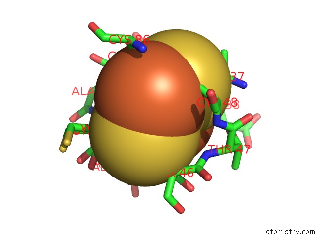
Mono view
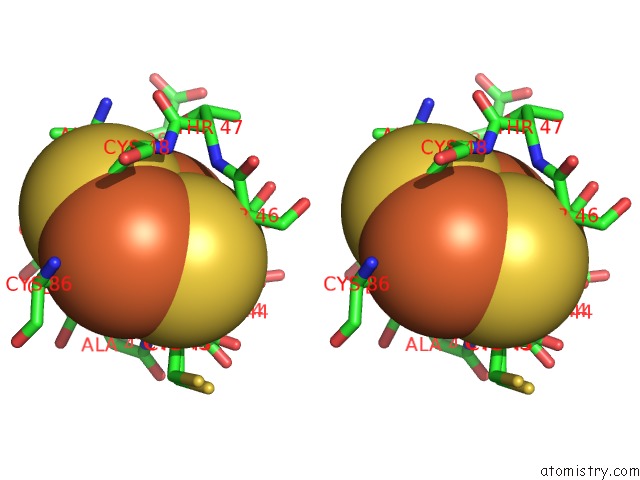
Stereo pair view
Mono view
Stereo pair view
A full contact list of Iron with other atoms in the Fe binding
site number 1 of Reduced Ferredoxin 6 From Rhodobacter Capsulatus within 5.0Å range:
|
Iron binding site 2 out of 2 in 1uwm
Go back to
Iron binding site 2 out
of 2 in the Reduced Ferredoxin 6 From Rhodobacter Capsulatus
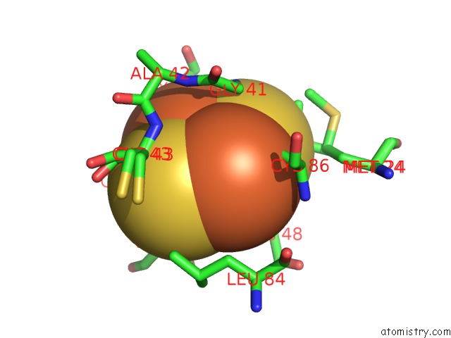
Mono view
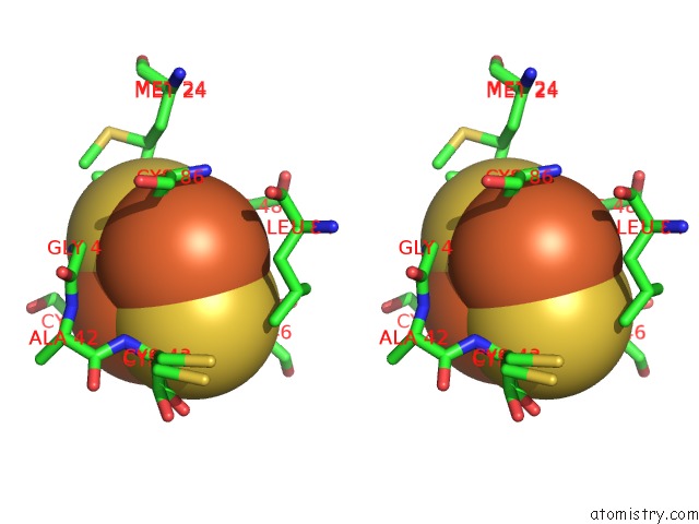
Stereo pair view
Mono view
Stereo pair view
A full contact list of Iron with other atoms in the Fe binding
site number 2 of Reduced Ferredoxin 6 From Rhodobacter Capsulatus within 5.0Å range:
|
Reference:
G.Sainz,
J.Jakoncic,
L.C.Sieker,
V.Stojanoff,
N.Sanishvili,
M.Asso,
P.Bertrand,
J.Armengaud,
Y.Jouanneau.
Structure of A [2FE-2S] Ferredoxin From Rhodobacter Capsulatus Likely Involved in Fe-S Cluster Biogenesis and Conformational Changes Observed Upon Reduction. J.Biol.Inorg.Chem. V. 11 235 2006.
ISSN: ISSN 0949-8257
PubMed: 16402206
DOI: 10.1007/S00775-005-0069-2
Page generated: Wed Jul 16 21:33:34 2025
ISSN: ISSN 0949-8257
PubMed: 16402206
DOI: 10.1007/S00775-005-0069-2
Last articles
Ni in 7ENHNi in 7ERR
Ni in 7EQV
Ni in 6ZJA
Ni in 7DH6
Ni in 7DGL
Ni in 7D2B
Ni in 7CPL
Ni in 7CPK
Ni in 7CXZ