Iron »
PDB 1uwm-1vme »
1v4w »
Iron in PDB 1v4w: Crystal Structure of Bluefin Tuna Hemoglobin Deoxy Form at PH7.5
Protein crystallography data
The structure of Crystal Structure of Bluefin Tuna Hemoglobin Deoxy Form at PH7.5, PDB code: 1v4w
was solved by
T.Yokoyama,
K.T.Chong,
Y.Miyazaki,
T.Nakatsukasa,
S.Unzai,
G.Miyazaki,
H.Morimoto,
R.H.T.Jeremy,
S.Y.Park,
with X-Ray Crystallography technique. A brief refinement statistics is given in the table below:
| Resolution Low / High (Å) | 13.84 / 1.70 |
| Space group | P 21 21 21 |
| Cell size a, b, c (Å), α, β, γ (°) | 56.766, 58.637, 174.414, 90.00, 90.00, 90.00 |
| R / Rfree (%) | 17.4 / 20.2 |
Iron Binding Sites:
The binding sites of Iron atom in the Crystal Structure of Bluefin Tuna Hemoglobin Deoxy Form at PH7.5
(pdb code 1v4w). This binding sites where shown within
5.0 Angstroms radius around Iron atom.
In total 4 binding sites of Iron where determined in the Crystal Structure of Bluefin Tuna Hemoglobin Deoxy Form at PH7.5, PDB code: 1v4w:
Jump to Iron binding site number: 1; 2; 3; 4;
In total 4 binding sites of Iron where determined in the Crystal Structure of Bluefin Tuna Hemoglobin Deoxy Form at PH7.5, PDB code: 1v4w:
Jump to Iron binding site number: 1; 2; 3; 4;
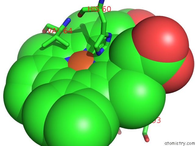
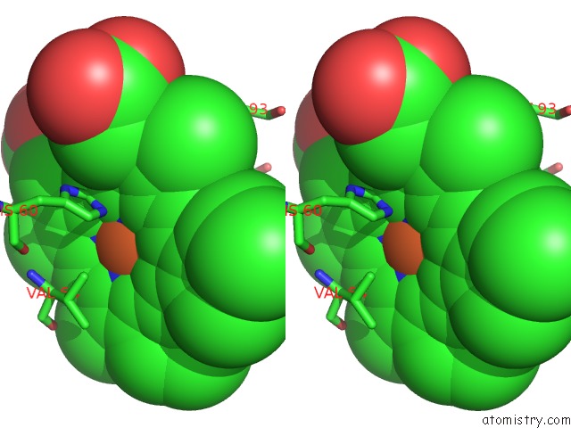
Iron binding site 1 out of 4 in 1v4w
Go back to
Iron binding site 1 out
of 4 in the Crystal Structure of Bluefin Tuna Hemoglobin Deoxy Form at PH7.5
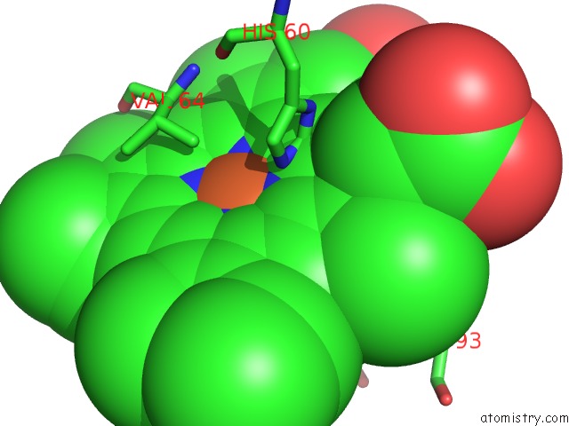
Mono view
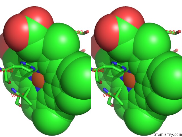
Stereo pair view
Mono view
Stereo pair view
A full contact list of Iron with other atoms in the Fe binding
site number 1 of Crystal Structure of Bluefin Tuna Hemoglobin Deoxy Form at PH7.5 within 5.0Å range:
|
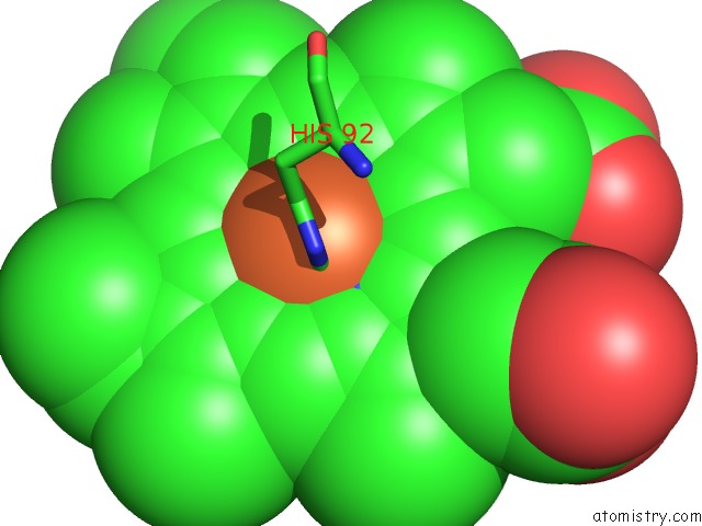
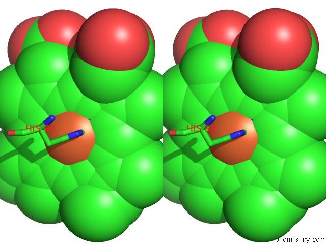
Iron binding site 2 out of 4 in 1v4w
Go back to
Iron binding site 2 out
of 4 in the Crystal Structure of Bluefin Tuna Hemoglobin Deoxy Form at PH7.5
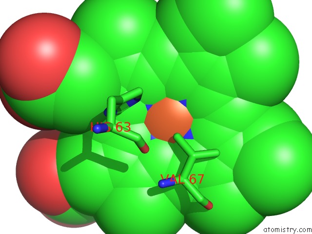
Mono view
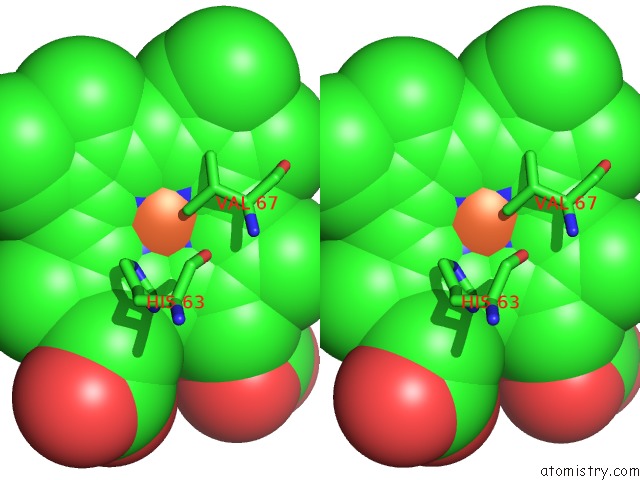
Stereo pair view
Mono view
Stereo pair view
A full contact list of Iron with other atoms in the Fe binding
site number 2 of Crystal Structure of Bluefin Tuna Hemoglobin Deoxy Form at PH7.5 within 5.0Å range:
|
Iron binding site 3 out of 4 in 1v4w
Go back to
Iron binding site 3 out
of 4 in the Crystal Structure of Bluefin Tuna Hemoglobin Deoxy Form at PH7.5
Mono view
Stereo pair view
Mono view
Stereo pair view
A full contact list of Iron with other atoms in the Fe binding
site number 3 of Crystal Structure of Bluefin Tuna Hemoglobin Deoxy Form at PH7.5 within 5.0Å range:
|
Iron binding site 4 out of 4 in 1v4w
Go back to
Iron binding site 4 out
of 4 in the Crystal Structure of Bluefin Tuna Hemoglobin Deoxy Form at PH7.5
Mono view
Stereo pair view
Mono view
Stereo pair view
A full contact list of Iron with other atoms in the Fe binding
site number 4 of Crystal Structure of Bluefin Tuna Hemoglobin Deoxy Form at PH7.5 within 5.0Å range:
|
Reference:
T.Yokoyama,
K.T.Chong,
G.Miyazaki,
H.Morimoto,
D.T.Shih,
S.Unzai,
J.R.Tame,
S.Y.Park.
Novel Mechanisms of pH Sensitivity in Tuna Hemoglobin: A Structural Explanation of the Root Effect J.Biol.Chem. V. 279 28632 2004.
ISSN: ISSN 0021-9258
PubMed: 15117955
DOI: 10.1074/JBC.M401740200
Page generated: Sat Aug 3 16:08:05 2024
ISSN: ISSN 0021-9258
PubMed: 15117955
DOI: 10.1074/JBC.M401740200
Last articles
Zn in 9MJ5Zn in 9HNW
Zn in 9G0L
Zn in 9FNE
Zn in 9DZN
Zn in 9E0I
Zn in 9D32
Zn in 9DAK
Zn in 8ZXC
Zn in 8ZUF