Iron »
PDB 1wa6-1x71 »
1wa6 »
Iron in PDB 1wa6: The Structure of Acc Oxidase
Enzymatic activity of The Structure of Acc Oxidase
All present enzymatic activity of The Structure of Acc Oxidase:
1.14.17.4;
1.14.17.4;
Protein crystallography data
The structure of The Structure of Acc Oxidase, PDB code: 1wa6
was solved by
Z.Zhang,
J.-S.Ren,
I.J.Clifton,
C.J.Schofield,
with X-Ray Crystallography technique. A brief refinement statistics is given in the table below:
| Resolution Low / High (Å) | 40.00 / 2.55 |
| Space group | I 2 2 2 |
| Cell size a, b, c (Å), α, β, γ (°) | 70.340, 107.060, 108.380, 90.00, 90.00, 90.00 |
| R / Rfree (%) | 18.6 / 23.6 |
Iron Binding Sites:
The binding sites of Iron atom in the The Structure of Acc Oxidase
(pdb code 1wa6). This binding sites where shown within
5.0 Angstroms radius around Iron atom.
In total only one binding site of Iron was determined in the The Structure of Acc Oxidase, PDB code: 1wa6:
In total only one binding site of Iron was determined in the The Structure of Acc Oxidase, PDB code: 1wa6:
Iron binding site 1 out of 1 in 1wa6
Go back to
Iron binding site 1 out
of 1 in the The Structure of Acc Oxidase
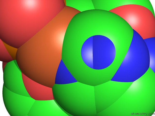
Mono view
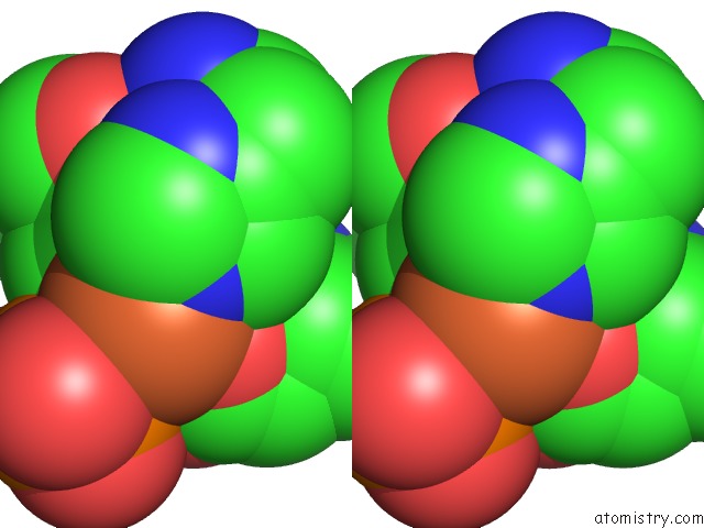
Stereo pair view
Mono view
Stereo pair view
A full contact list of Iron with other atoms in the Fe binding
site number 1 of The Structure of Acc Oxidase within 5.0Å range:
|
Reference:
Z.Zhang,
J.-S.Ren,
I.J.Clifton,
C.J.Schofield.
Crystal Structure and Mechanistic Implications of 1-Aminocyclopropane-1-Carboxylic Acid Oxidase (the Ethyling Forming Enzyme) Chem.Biol. V. 11 1383 2004.
ISSN: ISSN 1074-5521
PubMed: 15489165
DOI: 10.1016/J.CHEMBIOL.2004.08.012
Page generated: Wed Jul 16 21:49:44 2025
ISSN: ISSN 1074-5521
PubMed: 15489165
DOI: 10.1016/J.CHEMBIOL.2004.08.012
Last articles
Mn in 9LJUMn in 9LJW
Mn in 9LJS
Mn in 9LJR
Mn in 9LJT
Mn in 9LJV
Mg in 9UA2
Mg in 9R96
Mg in 9VM1
Mg in 9P01