Iron »
PDB 1x89-1xvc »
1x8n »
Iron in PDB 1x8n: 1.08 A Crystal Structure of Nitrophorin 4 From Rhodnius Prolixus Complexed with Nitric Oxide at pH 7.4
Protein crystallography data
The structure of 1.08 A Crystal Structure of Nitrophorin 4 From Rhodnius Prolixus Complexed with Nitric Oxide at pH 7.4, PDB code: 1x8n
was solved by
D.A.Kondrashov,
S.A.Roberts,
A.Weichsel,
W.R.Montfort,
with X-Ray Crystallography technique. A brief refinement statistics is given in the table below:
| Resolution Low / High (Å) | 6.00 / 1.08 |
| Space group | C 1 2 1 |
| Cell size a, b, c (Å), α, β, γ (°) | 70.147, 42.775, 53.042, 90.00, 94.09, 90.00 |
| R / Rfree (%) | 13.4 / 17.1 |
Iron Binding Sites:
The binding sites of Iron atom in the 1.08 A Crystal Structure of Nitrophorin 4 From Rhodnius Prolixus Complexed with Nitric Oxide at pH 7.4
(pdb code 1x8n). This binding sites where shown within
5.0 Angstroms radius around Iron atom.
In total only one binding site of Iron was determined in the 1.08 A Crystal Structure of Nitrophorin 4 From Rhodnius Prolixus Complexed with Nitric Oxide at pH 7.4, PDB code: 1x8n:
In total only one binding site of Iron was determined in the 1.08 A Crystal Structure of Nitrophorin 4 From Rhodnius Prolixus Complexed with Nitric Oxide at pH 7.4, PDB code: 1x8n:
Iron binding site 1 out of 1 in 1x8n
Go back to
Iron binding site 1 out
of 1 in the 1.08 A Crystal Structure of Nitrophorin 4 From Rhodnius Prolixus Complexed with Nitric Oxide at pH 7.4
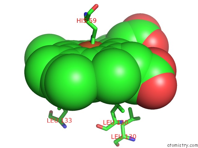
Mono view
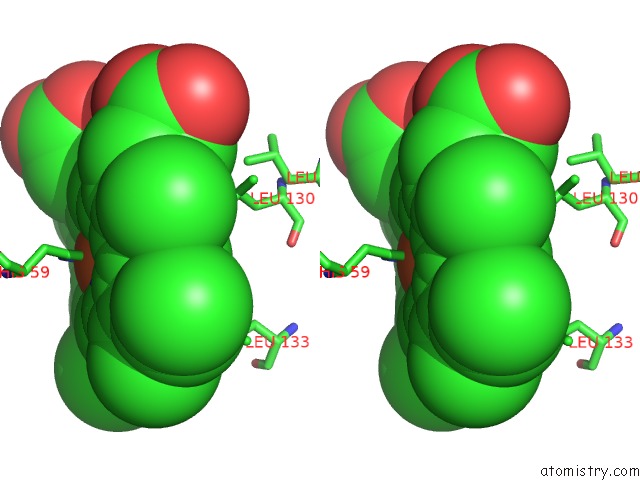
Stereo pair view
Mono view
Stereo pair view
A full contact list of Iron with other atoms in the Fe binding
site number 1 of 1.08 A Crystal Structure of Nitrophorin 4 From Rhodnius Prolixus Complexed with Nitric Oxide at pH 7.4 within 5.0Å range:
|
Reference:
D.A.Kondrashov,
S.A.Roberts,
A.Weichsel,
W.R.Montfort.
Protein Functional Cycle Viewed at Atomic Resolution: Conformational Change and Mobility in Nitrophorin 4 As A Function of pH and No Binding Biochemistry V. 43 13637 2004.
ISSN: ISSN 0006-2960
PubMed: 15504026
DOI: 10.1021/BI0483155
Page generated: Wed Jul 16 22:03:07 2025
ISSN: ISSN 0006-2960
PubMed: 15504026
DOI: 10.1021/BI0483155
Last articles
K in 5KOEK in 5KMT
K in 5KIL
K in 5KIK
K in 5KFX
K in 5KGR
K in 5KFW
K in 5KFV
K in 5KFU
K in 5KFT