Iron »
PDB 1xvd-1y4q »
1xvd »
Iron in PDB 1xvd: Soluble Methane Monooxygenase Hydroxylase: 4-Fluorophenol Soaked Structure
Enzymatic activity of Soluble Methane Monooxygenase Hydroxylase: 4-Fluorophenol Soaked Structure
All present enzymatic activity of Soluble Methane Monooxygenase Hydroxylase: 4-Fluorophenol Soaked Structure:
1.14.13.25;
1.14.13.25;
Protein crystallography data
The structure of Soluble Methane Monooxygenase Hydroxylase: 4-Fluorophenol Soaked Structure, PDB code: 1xvd
was solved by
M.H.Sazinsky,
S.J.Lippard,
with X-Ray Crystallography technique. A brief refinement statistics is given in the table below:
| Resolution Low / High (Å) | 29.94 / 2.30 |
| Space group | P 21 21 21 |
| Cell size a, b, c (Å), α, β, γ (°) | 71.182, 171.497, 221.304, 90.00, 90.00, 90.00 |
| R / Rfree (%) | 19.4 / 22.9 |
Other elements in 1xvd:
The structure of Soluble Methane Monooxygenase Hydroxylase: 4-Fluorophenol Soaked Structure also contains other interesting chemical elements:
| Fluorine | (F) | 2 atoms |
Iron Binding Sites:
The binding sites of Iron atom in the Soluble Methane Monooxygenase Hydroxylase: 4-Fluorophenol Soaked Structure
(pdb code 1xvd). This binding sites where shown within
5.0 Angstroms radius around Iron atom.
In total 4 binding sites of Iron where determined in the Soluble Methane Monooxygenase Hydroxylase: 4-Fluorophenol Soaked Structure, PDB code: 1xvd:
Jump to Iron binding site number: 1; 2; 3; 4;
In total 4 binding sites of Iron where determined in the Soluble Methane Monooxygenase Hydroxylase: 4-Fluorophenol Soaked Structure, PDB code: 1xvd:
Jump to Iron binding site number: 1; 2; 3; 4;
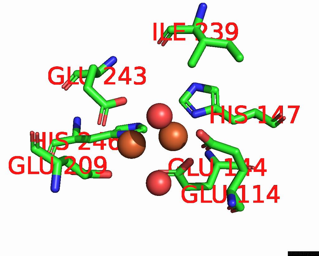
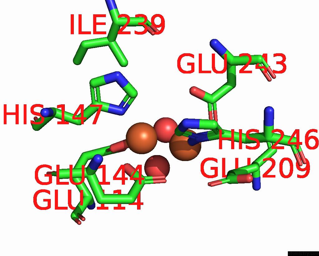
Iron binding site 1 out of 4 in 1xvd
Go back to
Iron binding site 1 out
of 4 in the Soluble Methane Monooxygenase Hydroxylase: 4-Fluorophenol Soaked Structure
Mono view
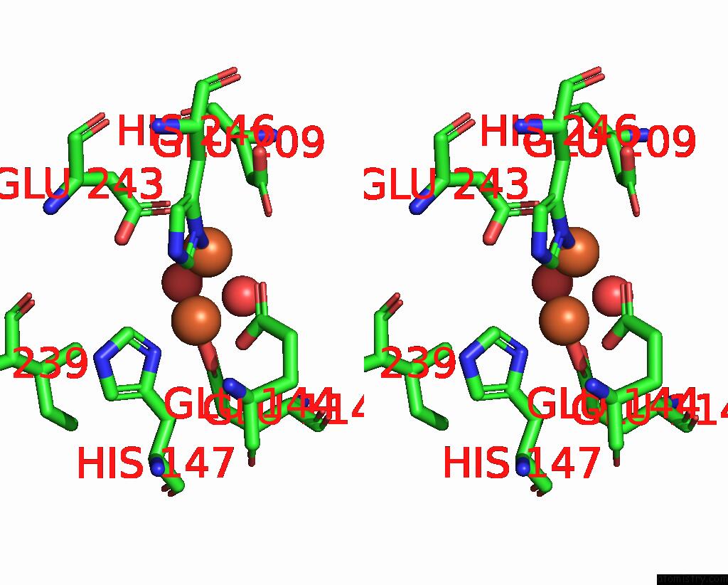
Stereo pair view
Mono view
Stereo pair view
A full contact list of Iron with other atoms in the Fe binding
site number 1 of Soluble Methane Monooxygenase Hydroxylase: 4-Fluorophenol Soaked Structure within 5.0Å range:
|
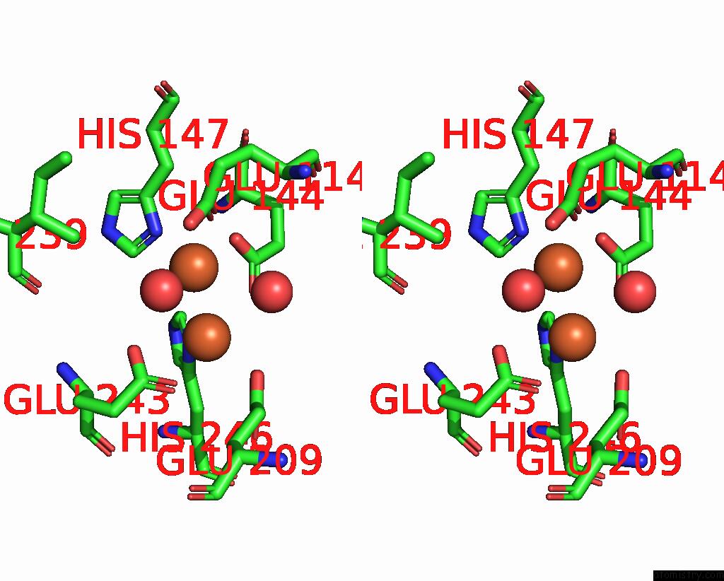
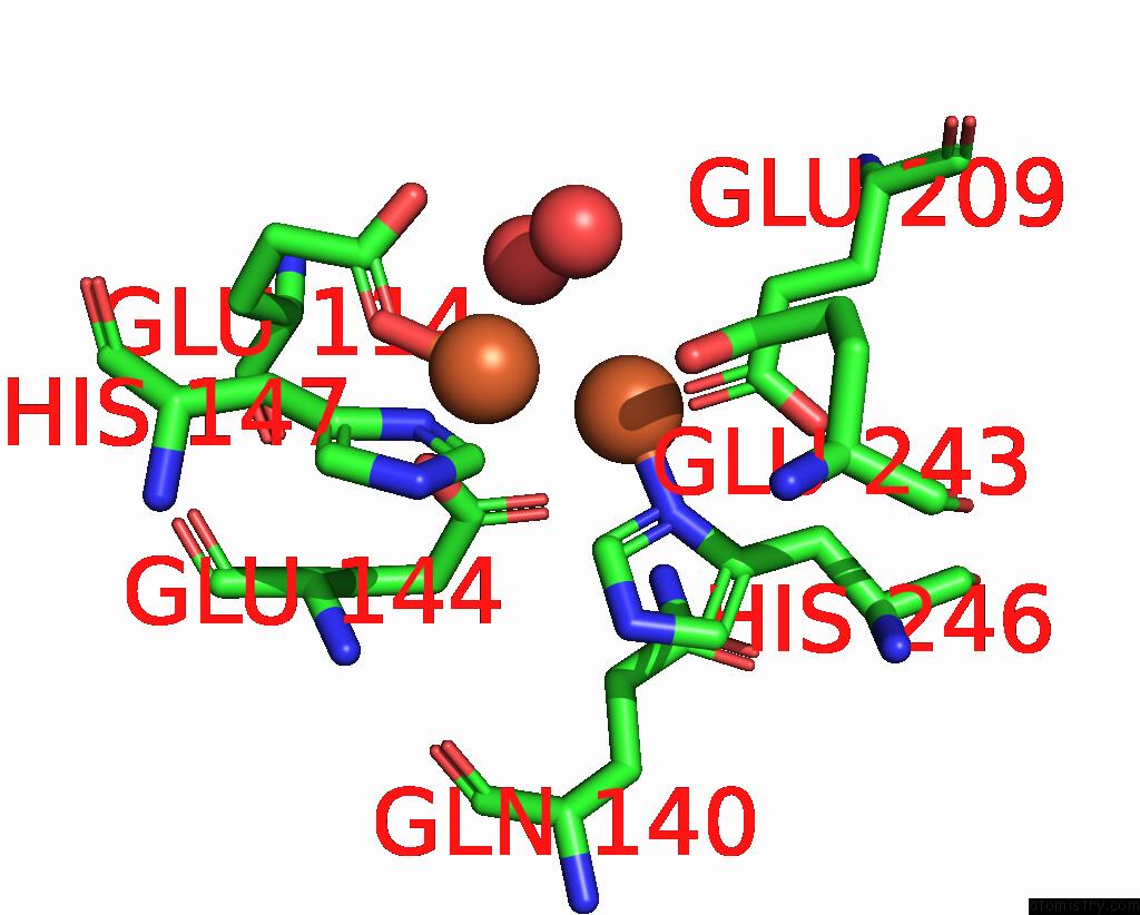
Iron binding site 2 out of 4 in 1xvd
Go back to
Iron binding site 2 out
of 4 in the Soluble Methane Monooxygenase Hydroxylase: 4-Fluorophenol Soaked Structure
Mono view
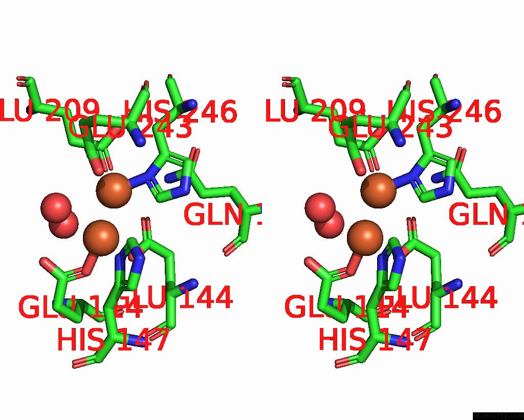
Stereo pair view
Mono view
Stereo pair view
A full contact list of Iron with other atoms in the Fe binding
site number 2 of Soluble Methane Monooxygenase Hydroxylase: 4-Fluorophenol Soaked Structure within 5.0Å range:
|
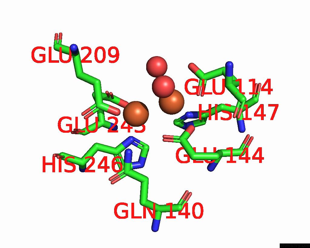
Iron binding site 3 out of 4 in 1xvd
Go back to
Iron binding site 3 out
of 4 in the Soluble Methane Monooxygenase Hydroxylase: 4-Fluorophenol Soaked Structure
Mono view
Stereo pair view
Mono view
Stereo pair view
A full contact list of Iron with other atoms in the Fe binding
site number 3 of Soluble Methane Monooxygenase Hydroxylase: 4-Fluorophenol Soaked Structure within 5.0Å range:
|
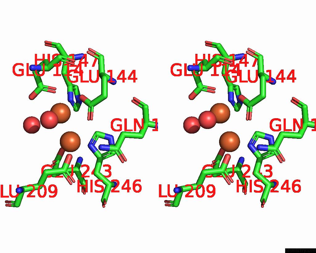
Iron binding site 4 out of 4 in 1xvd
Go back to
Iron binding site 4 out
of 4 in the Soluble Methane Monooxygenase Hydroxylase: 4-Fluorophenol Soaked Structure
Mono view
Stereo pair view
Mono view
Stereo pair view
A full contact list of Iron with other atoms in the Fe binding
site number 4 of Soluble Methane Monooxygenase Hydroxylase: 4-Fluorophenol Soaked Structure within 5.0Å range:
|
Reference:
M.H.Sazinsky,
S.J.Lippard.
Product Bound Structures of the Soluble Methane Monooxygenase Hydroxylase From Methylococcus Capsulatus (Bath): Protein Motion in the Alpha-Subunit J.Am.Chem.Soc. V. 127 5814 2005.
ISSN: ISSN 0002-7863
PubMed: 15839679
DOI: 10.1021/JA044099B
Page generated: Wed Jul 16 22:12:23 2025
ISSN: ISSN 0002-7863
PubMed: 15839679
DOI: 10.1021/JA044099B
Last articles
Mg in 3J7HMg in 3J7I
Mg in 3IVK
Mg in 3J6E
Mg in 3J6G
Mg in 3J6P
Mg in 3J6H
Mg in 3J1F
Mg in 3J6F
Mg in 3J5V