Iron »
PDB 1y4r-1yeu »
1ycb »
Iron in PDB 1ycb: Distal Pocket Polarity in Ligand Binding to Myoglobin: Deoxy and Carbonmonoxy Forms of A THREONINE68 (E11) Mutant Investigated By X-Ray Crystallography and Infrared Spectroscopy
Protein crystallography data
The structure of Distal Pocket Polarity in Ligand Binding to Myoglobin: Deoxy and Carbonmonoxy Forms of A THREONINE68 (E11) Mutant Investigated By X-Ray Crystallography and Infrared Spectroscopy, PDB code: 1ycb
was solved by
A.D.Cameron,
S.J.Smerdon,
A.J.Wilkinson,
J.Habash,
J.R.Helliwell,
with X-Ray Crystallography technique. A brief refinement statistics is given in the table below:
| Resolution Low / High (Å) | 8.00 / 2.10 |
| Space group | P 1 21 1 |
| Cell size a, b, c (Å), α, β, γ (°) | 124.600, 42.500, 92.000, 90.00, 92.00, 90.00 |
| R / Rfree (%) | n/a / n/a |
Iron Binding Sites:
The binding sites of Iron atom in the Distal Pocket Polarity in Ligand Binding to Myoglobin: Deoxy and Carbonmonoxy Forms of A THREONINE68 (E11) Mutant Investigated By X-Ray Crystallography and Infrared Spectroscopy
(pdb code 1ycb). This binding sites where shown within
5.0 Angstroms radius around Iron atom.
In total 2 binding sites of Iron where determined in the Distal Pocket Polarity in Ligand Binding to Myoglobin: Deoxy and Carbonmonoxy Forms of A THREONINE68 (E11) Mutant Investigated By X-Ray Crystallography and Infrared Spectroscopy, PDB code: 1ycb:
Jump to Iron binding site number: 1; 2;
In total 2 binding sites of Iron where determined in the Distal Pocket Polarity in Ligand Binding to Myoglobin: Deoxy and Carbonmonoxy Forms of A THREONINE68 (E11) Mutant Investigated By X-Ray Crystallography and Infrared Spectroscopy, PDB code: 1ycb:
Jump to Iron binding site number: 1; 2;
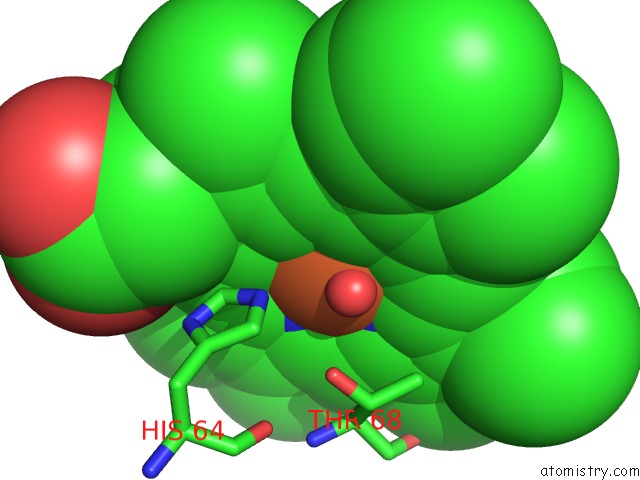
Iron binding site 1 out of 2 in 1ycb
Go back to
Iron binding site 1 out
of 2 in the Distal Pocket Polarity in Ligand Binding to Myoglobin: Deoxy and Carbonmonoxy Forms of A THREONINE68 (E11) Mutant Investigated By X-Ray Crystallography and Infrared Spectroscopy
Mono view
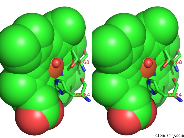
Stereo pair view
Mono view
Stereo pair view
A full contact list of Iron with other atoms in the Fe binding
site number 1 of Distal Pocket Polarity in Ligand Binding to Myoglobin: Deoxy and Carbonmonoxy Forms of A THREONINE68 (E11) Mutant Investigated By X-Ray Crystallography and Infrared Spectroscopy within 5.0Å range:
|
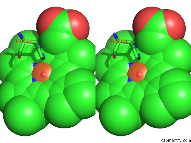
Iron binding site 2 out of 2 in 1ycb
Go back to
Iron binding site 2 out
of 2 in the Distal Pocket Polarity in Ligand Binding to Myoglobin: Deoxy and Carbonmonoxy Forms of A THREONINE68 (E11) Mutant Investigated By X-Ray Crystallography and Infrared Spectroscopy
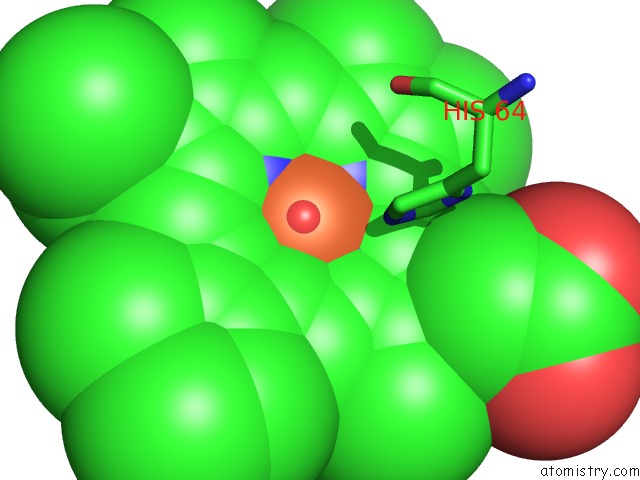
Mono view
Stereo pair view
Mono view
Stereo pair view
A full contact list of Iron with other atoms in the Fe binding
site number 2 of Distal Pocket Polarity in Ligand Binding to Myoglobin: Deoxy and Carbonmonoxy Forms of A THREONINE68 (E11) Mutant Investigated By X-Ray Crystallography and Infrared Spectroscopy within 5.0Å range:
|
Reference:
A.D.Cameron,
S.J.Smerdon,
A.J.Wilkinson,
J.Habash,
J.R.Helliwell,
T.Li,
J.S.Olson.
Distal Pocket Polarity in Ligand Binding to Myoglobin: Deoxy and Carbonmonoxy Forms of A THREONINE68(E11) Mutant Investigated By X-Ray Crystallography and Infrared Spectroscopy. Biochemistry V. 32 13061 1993.
ISSN: ISSN 0006-2960
PubMed: 8241160
DOI: 10.1021/BI00211A016
Page generated: Wed Jul 16 22:34:52 2025
ISSN: ISSN 0006-2960
PubMed: 8241160
DOI: 10.1021/BI00211A016
Last articles
K in 6WP3K in 6WLV
K in 6WMG
K in 6WMF
K in 6WME
K in 6WH5
K in 6WIC
K in 6WFL
K in 6WDY
K in 6WDX