Iron »
PDB 1yev-1yqo »
1yma »
Iron in PDB 1yma: Structural Characterization of Heme Ligation in the HIS64-->Tyr Variant of Myoglobin
Protein crystallography data
The structure of Structural Characterization of Heme Ligation in the HIS64-->Tyr Variant of Myoglobin, PDB code: 1yma
was solved by
R.Maurus,
G.D.Brayer,
with X-Ray Crystallography technique. A brief refinement statistics is given in the table below:
| Resolution Low / High (Å) | 8.00 / 2.00 |
| Space group | P 1 21 1 |
| Cell size a, b, c (Å), α, β, γ (°) | 63.700, 28.800, 35.700, 90.00, 106.60, 90.00 |
| R / Rfree (%) | n/a / n/a |
Iron Binding Sites:
The binding sites of Iron atom in the Structural Characterization of Heme Ligation in the HIS64-->Tyr Variant of Myoglobin
(pdb code 1yma). This binding sites where shown within
5.0 Angstroms radius around Iron atom.
In total only one binding site of Iron was determined in the Structural Characterization of Heme Ligation in the HIS64-->Tyr Variant of Myoglobin, PDB code: 1yma:
In total only one binding site of Iron was determined in the Structural Characterization of Heme Ligation in the HIS64-->Tyr Variant of Myoglobin, PDB code: 1yma:
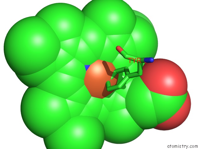
Iron binding site 1 out of 1 in 1yma
Go back to
Iron binding site 1 out
of 1 in the Structural Characterization of Heme Ligation in the HIS64-->Tyr Variant of Myoglobin
Mono view

Stereo pair view
Mono view
Stereo pair view
A full contact list of Iron with other atoms in the Fe binding
site number 1 of Structural Characterization of Heme Ligation in the HIS64-->Tyr Variant of Myoglobin within 5.0Å range:
|
Reference:
R.Maurus,
R.Bogumil,
Y.Luo,
H.L.Tang,
M.Smith,
A.G.Mauk,
G.D.Brayer.
Structural Characterization of Heme Ligation in the HIS64-->Tyr Variant of Myoglobin. J.Biol.Chem. V. 269 12606 1994.
ISSN: ISSN 0021-9258
PubMed: 8175669
Page generated: Wed Jul 16 22:48:26 2025
ISSN: ISSN 0021-9258
PubMed: 8175669
Last articles
Mg in 4EMWMg in 4ELT
Mg in 4ELU
Mg in 4ELV
Mg in 4EM3
Mg in 4EKD
Mg in 4EHU
Mg in 4EHT
Mg in 4EKC
Mg in 4EHY