Iron »
PDB 1yqp-1zj9 »
1yzp »
Iron in PDB 1yzp: Substrate-Free Manganese Peroxidase
Enzymatic activity of Substrate-Free Manganese Peroxidase
All present enzymatic activity of Substrate-Free Manganese Peroxidase:
1.11.1.13;
1.11.1.13;
Protein crystallography data
The structure of Substrate-Free Manganese Peroxidase, PDB code: 1yzp
was solved by
M.Sundaramoorthy,
H.L.Youngs,
M.H.Gold,
T.L.Poulos,
with X-Ray Crystallography technique. A brief refinement statistics is given in the table below:
| Resolution Low / High (Å) | 8.00 / 1.60 |
| Space group | C 1 2 1 |
| Cell size a, b, c (Å), α, β, γ (°) | 160.595, 45.356, 52.817, 90.00, 97.25, 90.00 |
| R / Rfree (%) | 15.3 / 20.3 |
Other elements in 1yzp:
The structure of Substrate-Free Manganese Peroxidase also contains other interesting chemical elements:
| Calcium | (Ca) | 2 atoms |
Iron Binding Sites:
The binding sites of Iron atom in the Substrate-Free Manganese Peroxidase
(pdb code 1yzp). This binding sites where shown within
5.0 Angstroms radius around Iron atom.
In total only one binding site of Iron was determined in the Substrate-Free Manganese Peroxidase, PDB code: 1yzp:
In total only one binding site of Iron was determined in the Substrate-Free Manganese Peroxidase, PDB code: 1yzp:
Iron binding site 1 out of 1 in 1yzp
Go back to
Iron binding site 1 out
of 1 in the Substrate-Free Manganese Peroxidase
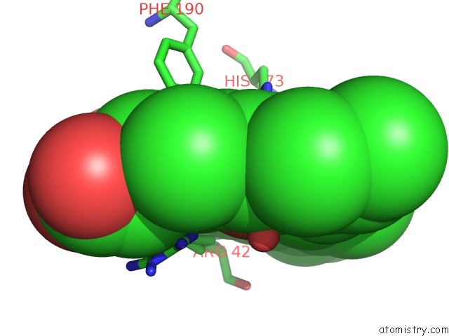
Mono view
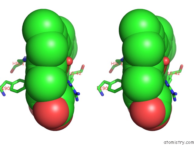
Stereo pair view
Mono view
Stereo pair view
A full contact list of Iron with other atoms in the Fe binding
site number 1 of Substrate-Free Manganese Peroxidase within 5.0Å range:
|
Reference:
M.Sundaramoorthy,
H.L.Youngs,
M.H.Gold,
T.L.Poulos.
High-Resolution Crystal Structure of Manganese Peroxidase: Substrate and Inhibitor Complexes. Biochemistry V. 44 6463 2005.
ISSN: ISSN 0006-2960
PubMed: 15850380
DOI: 10.1021/BI047318E
Page generated: Wed Jul 16 22:58:42 2025
ISSN: ISSN 0006-2960
PubMed: 15850380
DOI: 10.1021/BI047318E
Last articles
Mg in 5MM7Mg in 5ML7
Mg in 5MM9
Mg in 5MLB
Mg in 5MM4
Mg in 5MLZ
Mg in 5MLA
Mg in 5MIL
Mg in 5MIO
Mg in 5MHA