Iron »
PDB 1zlq-2ai5 »
2acp »
Iron in PDB 2acp: Crystal Structure of Nitrophorin 2 Aqua Complex
Protein crystallography data
The structure of Crystal Structure of Nitrophorin 2 Aqua Complex, PDB code: 2acp
was solved by
A.Weichsel,
R.E.Berry,
F.A.Walker,
W.R.Montfort,
with X-Ray Crystallography technique. A brief refinement statistics is given in the table below:
| Resolution Low / High (Å) | 20.00 / 1.40 |
| Space group | P 21 21 2 |
| Cell size a, b, c (Å), α, β, γ (°) | 39.754, 125.642, 33.468, 90.00, 90.00, 90.00 |
| R / Rfree (%) | 18.5 / 23.3 |
Iron Binding Sites:
The binding sites of Iron atom in the Crystal Structure of Nitrophorin 2 Aqua Complex
(pdb code 2acp). This binding sites where shown within
5.0 Angstroms radius around Iron atom.
In total only one binding site of Iron was determined in the Crystal Structure of Nitrophorin 2 Aqua Complex, PDB code: 2acp:
In total only one binding site of Iron was determined in the Crystal Structure of Nitrophorin 2 Aqua Complex, PDB code: 2acp:
Iron binding site 1 out of 1 in 2acp
Go back to
Iron binding site 1 out
of 1 in the Crystal Structure of Nitrophorin 2 Aqua Complex
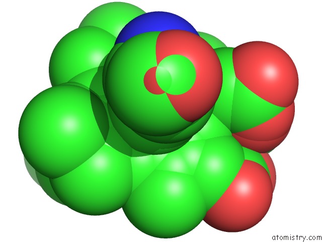
Mono view
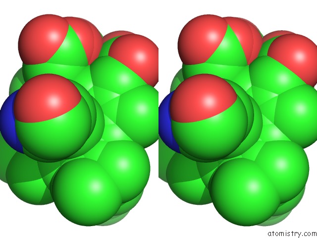
Stereo pair view
Mono view
Stereo pair view
A full contact list of Iron with other atoms in the Fe binding
site number 1 of Crystal Structure of Nitrophorin 2 Aqua Complex within 5.0Å range:
|
Reference:
A.Weichsel,
R.E.Berry,
F.A.Walker,
W.R.Montfort.
Crystal Structures, Ligand Induced Conformational Change and Heme Deformation in Complexes of Nitrophorin 2, A Nitric Oxide Transport Protein From Rhodnius Prolixus To Be Published.
Page generated: Wed Jul 16 23:26:06 2025
Last articles
Na in 4C79Na in 4C76
Na in 4C6Y
Na in 4C6S
Na in 4C75
Na in 4C6W
Na in 4C3Y
Na in 4C3X
Na in 4C3V
Na in 4C44