Iron »
PDB 2ays-2bmm »
2azq »
Iron in PDB 2azq: Crystal Structure of Catechol 1,2-Dioxygenase From Pseudomonas Arvilla C-1
Enzymatic activity of Crystal Structure of Catechol 1,2-Dioxygenase From Pseudomonas Arvilla C-1
All present enzymatic activity of Crystal Structure of Catechol 1,2-Dioxygenase From Pseudomonas Arvilla C-1:
1.13.11.1;
1.13.11.1;
Protein crystallography data
The structure of Crystal Structure of Catechol 1,2-Dioxygenase From Pseudomonas Arvilla C-1, PDB code: 2azq
was solved by
C.A.Earhart,
M.W.Vetting,
R.Gosu,
I.Michaud-Soret,
L.Que,
D.H.Ohlendorf,
with X-Ray Crystallography technique. A brief refinement statistics is given in the table below:
| Resolution Low / High (Å) | 21.00 / 2.65 |
| Space group | C 2 2 21 |
| Cell size a, b, c (Å), α, β, γ (°) | 62.730, 71.520, 187.090, 90.00, 90.00, 90.00 |
| R / Rfree (%) | 22.9 / 28.4 |
Iron Binding Sites:
The binding sites of Iron atom in the Crystal Structure of Catechol 1,2-Dioxygenase From Pseudomonas Arvilla C-1
(pdb code 2azq). This binding sites where shown within
5.0 Angstroms radius around Iron atom.
In total only one binding site of Iron was determined in the Crystal Structure of Catechol 1,2-Dioxygenase From Pseudomonas Arvilla C-1, PDB code: 2azq:
In total only one binding site of Iron was determined in the Crystal Structure of Catechol 1,2-Dioxygenase From Pseudomonas Arvilla C-1, PDB code: 2azq:
Iron binding site 1 out of 1 in 2azq
Go back to
Iron binding site 1 out
of 1 in the Crystal Structure of Catechol 1,2-Dioxygenase From Pseudomonas Arvilla C-1
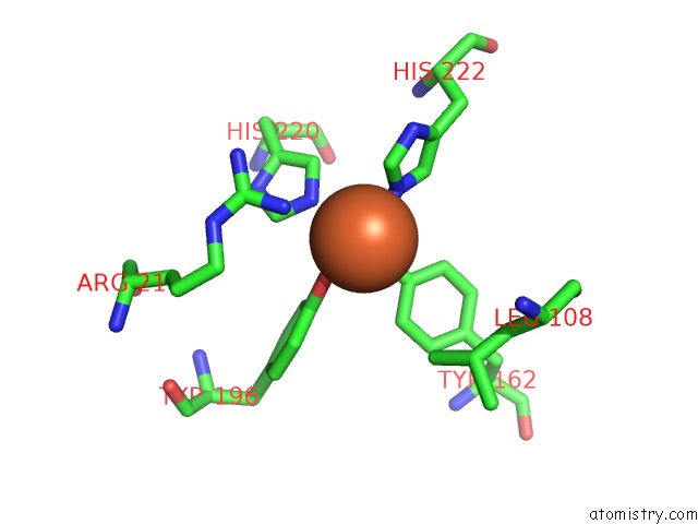
Mono view
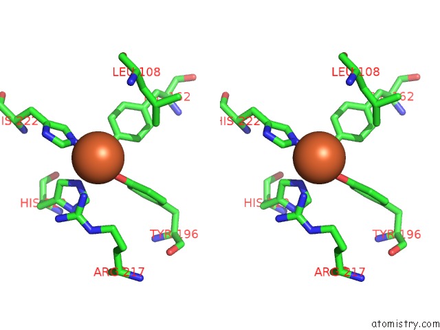
Stereo pair view
Mono view
Stereo pair view
A full contact list of Iron with other atoms in the Fe binding
site number 1 of Crystal Structure of Catechol 1,2-Dioxygenase From Pseudomonas Arvilla C-1 within 5.0Å range:
|
Reference:
C.A.Earhart,
M.W.Vetting,
R.Gosu,
I.Michaud-Soret,
L.Que,
D.H.Ohlendorf.
Structure of Catechol 1,2-Dioxygenase From Pseudomonas Arvilla Biochem.Biophys.Res.Commun. V. 338 198 2005.
ISSN: ISSN 0006-291X
PubMed: 16171781
DOI: 10.1016/J.BBRC.2005.08.221
Page generated: Wed Jul 16 23:46:51 2025
ISSN: ISSN 0006-291X
PubMed: 16171781
DOI: 10.1016/J.BBRC.2005.08.221
Last articles
Mg in 4YN0Mg in 4YMU
Mg in 4YMN
Mg in 4YLP
Mg in 4YMG
Mg in 4YLO
Mg in 4YLN
Mg in 4YLG
Mg in 4YKH
Mg in 4YKP