Iron »
PDB 2ays-2bmm »
2bkm »
Iron in PDB 2bkm: Crystal Structure of the Truncated Hemoglobin From Geobacillus Stearothermophilus
Protein crystallography data
The structure of Crystal Structure of the Truncated Hemoglobin From Geobacillus Stearothermophilus, PDB code: 2bkm
was solved by
A.Ilari,
P.Kjelgaard,
C.Von Wachenfeldt,
A.Boffi,
E.Chiancone,
with X-Ray Crystallography technique. A brief refinement statistics is given in the table below:
| Resolution Low / High (Å) | 25.00 / 1.50 |
| Space group | P 1 21 1 |
| Cell size a, b, c (Å), α, β, γ (°) | 35.123, 99.339, 35.252, 90.00, 105.59, 90.00 |
| R / Rfree (%) | 16.7 / 19.4 |
Iron Binding Sites:
The binding sites of Iron atom in the Crystal Structure of the Truncated Hemoglobin From Geobacillus Stearothermophilus
(pdb code 2bkm). This binding sites where shown within
5.0 Angstroms radius around Iron atom.
In total 2 binding sites of Iron where determined in the Crystal Structure of the Truncated Hemoglobin From Geobacillus Stearothermophilus, PDB code: 2bkm:
Jump to Iron binding site number: 1; 2;
In total 2 binding sites of Iron where determined in the Crystal Structure of the Truncated Hemoglobin From Geobacillus Stearothermophilus, PDB code: 2bkm:
Jump to Iron binding site number: 1; 2;
Iron binding site 1 out of 2 in 2bkm
Go back to
Iron binding site 1 out
of 2 in the Crystal Structure of the Truncated Hemoglobin From Geobacillus Stearothermophilus

Mono view
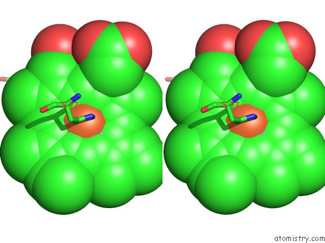
Stereo pair view
Mono view
Stereo pair view
A full contact list of Iron with other atoms in the Fe binding
site number 1 of Crystal Structure of the Truncated Hemoglobin From Geobacillus Stearothermophilus within 5.0Å range:
|
Iron binding site 2 out of 2 in 2bkm
Go back to
Iron binding site 2 out
of 2 in the Crystal Structure of the Truncated Hemoglobin From Geobacillus Stearothermophilus
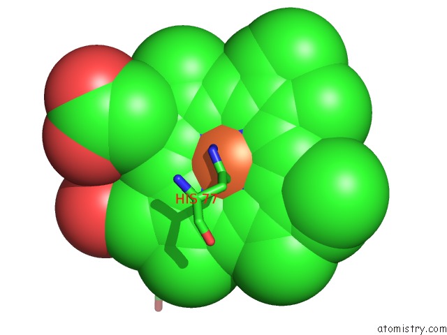
Mono view

Stereo pair view
Mono view
Stereo pair view
A full contact list of Iron with other atoms in the Fe binding
site number 2 of Crystal Structure of the Truncated Hemoglobin From Geobacillus Stearothermophilus within 5.0Å range:
|
Reference:
A.Ilari,
P.Kjelgaard,
C.Von Wachenfeldt,
B.Catacchio,
E.Chiancone,
A.Boffi.
Crystal Structure and Ligand Binding Properties of the Truncated Hemoglobin From Geobacillus Stearothermophilus Arch.Biochem.Biophys. V. 457 85 2007.
ISSN: ISSN 0003-9861
PubMed: 17126283
DOI: 10.1016/J.ABB.2006.09.033
Page generated: Wed Jul 16 23:54:47 2025
ISSN: ISSN 0003-9861
PubMed: 17126283
DOI: 10.1016/J.ABB.2006.09.033
Last articles
Mg in 3J7HMg in 3J7I
Mg in 3IVK
Mg in 3J6E
Mg in 3J6G
Mg in 3J6P
Mg in 3J6H
Mg in 3J1F
Mg in 3J6F
Mg in 3J5V