Iron »
PDB 2bmo-2c1v »
2bum »
Iron in PDB 2bum: Crystal Structure of Wild-Type Protocatechuate 3,4-Dioxygenase From Acinetobacter Sp. ADP1
Enzymatic activity of Crystal Structure of Wild-Type Protocatechuate 3,4-Dioxygenase From Acinetobacter Sp. ADP1
All present enzymatic activity of Crystal Structure of Wild-Type Protocatechuate 3,4-Dioxygenase From Acinetobacter Sp. ADP1:
1.13.11.3;
1.13.11.3;
Protein crystallography data
The structure of Crystal Structure of Wild-Type Protocatechuate 3,4-Dioxygenase From Acinetobacter Sp. ADP1, PDB code: 2bum
was solved by
M.W.Vetting,
M.P.Valley,
D.A.D'argenio,
L.N.Ornston,
J.D.Lipscomb,
D.H.Ohlendorf,
with X-Ray Crystallography technique. A brief refinement statistics is given in the table below:
| Resolution Low / High (Å) | 20.00 / 1.80 |
| Space group | I 2 3 |
| Cell size a, b, c (Å), α, β, γ (°) | 145.230, 145.230, 145.230, 90.00, 90.00, 90.00 |
| R / Rfree (%) | 18.5 / 21 |
Iron Binding Sites:
The binding sites of Iron atom in the Crystal Structure of Wild-Type Protocatechuate 3,4-Dioxygenase From Acinetobacter Sp. ADP1
(pdb code 2bum). This binding sites where shown within
5.0 Angstroms radius around Iron atom.
In total only one binding site of Iron was determined in the Crystal Structure of Wild-Type Protocatechuate 3,4-Dioxygenase From Acinetobacter Sp. ADP1, PDB code: 2bum:
In total only one binding site of Iron was determined in the Crystal Structure of Wild-Type Protocatechuate 3,4-Dioxygenase From Acinetobacter Sp. ADP1, PDB code: 2bum:
Iron binding site 1 out of 1 in 2bum
Go back to
Iron binding site 1 out
of 1 in the Crystal Structure of Wild-Type Protocatechuate 3,4-Dioxygenase From Acinetobacter Sp. ADP1

Mono view
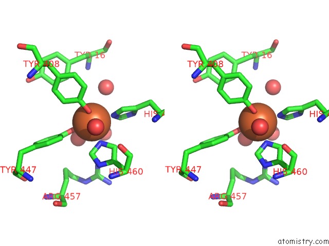
Stereo pair view
Mono view
Stereo pair view
A full contact list of Iron with other atoms in the Fe binding
site number 1 of Crystal Structure of Wild-Type Protocatechuate 3,4-Dioxygenase From Acinetobacter Sp. ADP1 within 5.0Å range:
|
Reference:
C.K.Brown,
M.W.Vetting,
C.A.Earhart,
D.H.Ohlendorf.
Biophysical Analyses of Designed and Selected Mutants of Protocatechuate 3,4-Dioxygenase Annu.Rev.Microbiol. V. 58 555 2004.
ISSN: ISSN 0066-4227
PubMed: 15487948
DOI: 10.1146/ANNUREV.MICRO.57.030502.090927
Page generated: Thu Jul 17 00:01:58 2025
ISSN: ISSN 0066-4227
PubMed: 15487948
DOI: 10.1146/ANNUREV.MICRO.57.030502.090927
Last articles
Mn in 9LJUMn in 9LJW
Mn in 9LJS
Mn in 9LJR
Mn in 9LJT
Mn in 9LJV
Mg in 9UA2
Mg in 9R96
Mg in 9VM1
Mg in 9P01