Iron »
PDB 2ciz-2d3q »
2cmm »
Iron in PDB 2cmm: Structural Analysis of the Myoglobin Reconstituted with Iron Porphine
Protein crystallography data
The structure of Structural Analysis of the Myoglobin Reconstituted with Iron Porphine, PDB code: 2cmm
was solved by
T.Sato,
N.Tanaka,
H.Moriyama,
N.Igarashi,
S.Neya,
N.Funasaki,
T.Iizuka,
Y.Shiro,
with X-Ray Crystallography technique. A brief refinement statistics is given in the table below:
| Resolution Low / High (Å) | N/A / 1.80 |
| Space group | P 21 21 21 |
| Cell size a, b, c (Å), α, β, γ (°) | 57.620, 76.230, 33.260, 90.00, 90.00, 90.00 |
| R / Rfree (%) | n/a / n/a |
Iron Binding Sites:
The binding sites of Iron atom in the Structural Analysis of the Myoglobin Reconstituted with Iron Porphine
(pdb code 2cmm). This binding sites where shown within
5.0 Angstroms radius around Iron atom.
In total only one binding site of Iron was determined in the Structural Analysis of the Myoglobin Reconstituted with Iron Porphine, PDB code: 2cmm:
In total only one binding site of Iron was determined in the Structural Analysis of the Myoglobin Reconstituted with Iron Porphine, PDB code: 2cmm:
Iron binding site 1 out of 1 in 2cmm
Go back to
Iron binding site 1 out
of 1 in the Structural Analysis of the Myoglobin Reconstituted with Iron Porphine
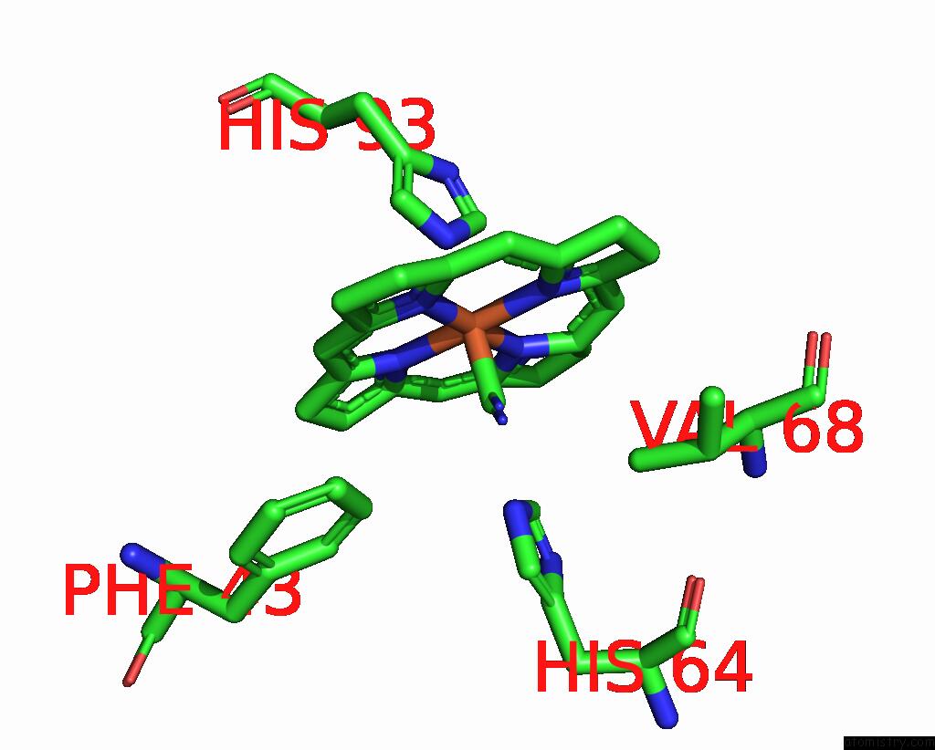
Mono view
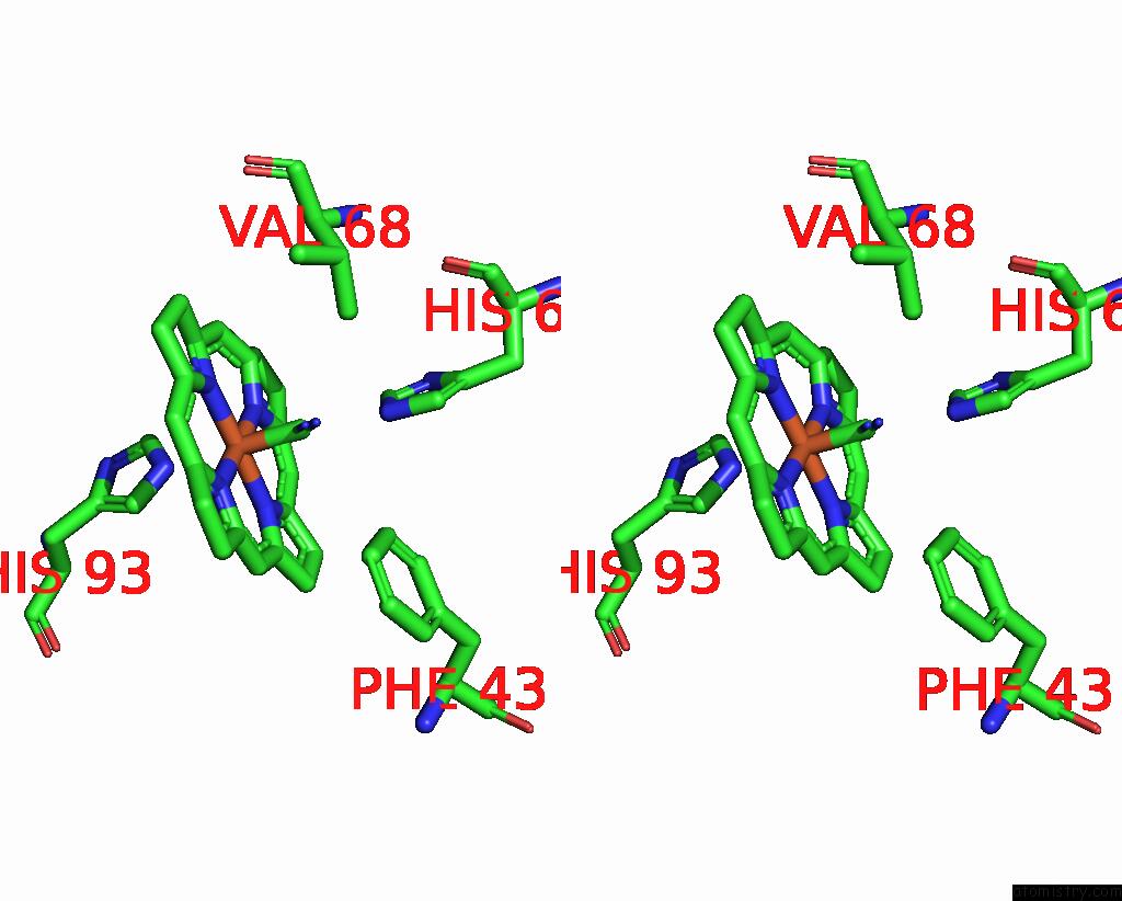
Stereo pair view
Mono view
Stereo pair view
A full contact list of Iron with other atoms in the Fe binding
site number 1 of Structural Analysis of the Myoglobin Reconstituted with Iron Porphine within 5.0Å range:
|
Reference:
S.Neya,
N.Funasaki,
T.Sato,
N.Igarashi,
N.Tanaka.
Structural Analysis of the Myoglobin Reconstituted with Iron Porphine. J.Biol.Chem. V. 268 8935 1993.
ISSN: ISSN 0021-9258
PubMed: 8473336
Page generated: Thu Jul 17 00:28:25 2025
ISSN: ISSN 0021-9258
PubMed: 8473336
Last articles
Na in 3MICNa in 3MI3
Na in 3MIE
Na in 3MGI
Na in 3ME4
Na in 3MDC
Na in 3MGA
Na in 3MEQ
Na in 3ME2
Na in 3MDA