Iron »
PDB 2ciz-2d3q »
2cn4 »
Iron in PDB 2cn4: The Crystal Structure of the Secreted Dimeric Form of the Hemophore Hasa Reveals A Domain Swapping with An Exchanged Heme Ligand
Protein crystallography data
The structure of The Crystal Structure of the Secreted Dimeric Form of the Hemophore Hasa Reveals A Domain Swapping with An Exchanged Heme Ligand, PDB code: 2cn4
was solved by
M.Czjzek,
S.Letoffe,
C.Wandersman,
M.Delepierre,
A.Lecroisey,
N.Izadi-Pruneyre,
with X-Ray Crystallography technique. A brief refinement statistics is given in the table below:
| Resolution Low / High (Å) | 30.00 / 2.3 |
| Space group | P 1 21 1 |
| Cell size a, b, c (Å), α, β, γ (°) | 47.080, 62.320, 56.820, 90.00, 105.34, 90.00 |
| R / Rfree (%) | 16.9 / 22.4 |
Iron Binding Sites:
The binding sites of Iron atom in the The Crystal Structure of the Secreted Dimeric Form of the Hemophore Hasa Reveals A Domain Swapping with An Exchanged Heme Ligand
(pdb code 2cn4). This binding sites where shown within
5.0 Angstroms radius around Iron atom.
In total 2 binding sites of Iron where determined in the The Crystal Structure of the Secreted Dimeric Form of the Hemophore Hasa Reveals A Domain Swapping with An Exchanged Heme Ligand, PDB code: 2cn4:
Jump to Iron binding site number: 1; 2;
In total 2 binding sites of Iron where determined in the The Crystal Structure of the Secreted Dimeric Form of the Hemophore Hasa Reveals A Domain Swapping with An Exchanged Heme Ligand, PDB code: 2cn4:
Jump to Iron binding site number: 1; 2;
Iron binding site 1 out of 2 in 2cn4
Go back to
Iron binding site 1 out
of 2 in the The Crystal Structure of the Secreted Dimeric Form of the Hemophore Hasa Reveals A Domain Swapping with An Exchanged Heme Ligand
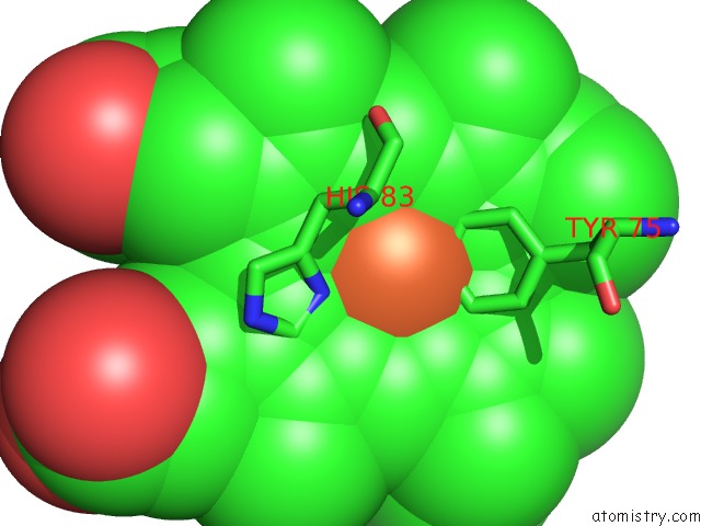
Mono view
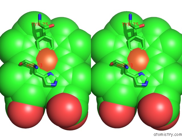
Stereo pair view
Mono view
Stereo pair view
A full contact list of Iron with other atoms in the Fe binding
site number 1 of The Crystal Structure of the Secreted Dimeric Form of the Hemophore Hasa Reveals A Domain Swapping with An Exchanged Heme Ligand within 5.0Å range:
|
Iron binding site 2 out of 2 in 2cn4
Go back to
Iron binding site 2 out
of 2 in the The Crystal Structure of the Secreted Dimeric Form of the Hemophore Hasa Reveals A Domain Swapping with An Exchanged Heme Ligand
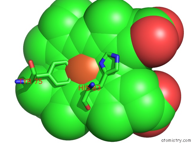
Mono view
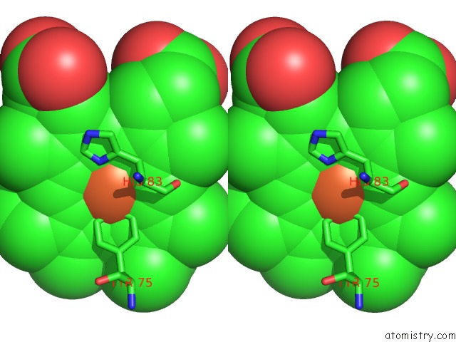
Stereo pair view
Mono view
Stereo pair view
A full contact list of Iron with other atoms in the Fe binding
site number 2 of The Crystal Structure of the Secreted Dimeric Form of the Hemophore Hasa Reveals A Domain Swapping with An Exchanged Heme Ligand within 5.0Å range:
|
Reference:
M.Czjzek,
S.Letoffe,
C.Wandersman,
M.Delepierre,
A.Lecroisey,
N.Izadi-Pruneyre.
The Crystal Structure of the Secreted Dimeric Form of the Hemophore Hasa Reveals A Domain Swapping with An Exchanged Heme Ligand J.Mol.Biol. V. 365 1176 2007.
ISSN: ISSN 0022-2836
PubMed: 17113104
DOI: 10.1016/J.JMB.2006.10.063
Page generated: Thu Jul 17 00:28:59 2025
ISSN: ISSN 0022-2836
PubMed: 17113104
DOI: 10.1016/J.JMB.2006.10.063
Last articles
Fe in 2YXOFe in 2YRS
Fe in 2YXC
Fe in 2YNM
Fe in 2YVJ
Fe in 2YP1
Fe in 2YU2
Fe in 2YU1
Fe in 2YQB
Fe in 2YOO