Iron »
PDB 2ciz-2d3q »
2cw2 »
Iron in PDB 2cw2: Crystal Structure of Superoxide Dismutase From P. Marinus
Enzymatic activity of Crystal Structure of Superoxide Dismutase From P. Marinus
All present enzymatic activity of Crystal Structure of Superoxide Dismutase From P. Marinus:
1.15.1.1;
1.15.1.1;
Protein crystallography data
The structure of Crystal Structure of Superoxide Dismutase From P. Marinus, PDB code: 2cw2
was solved by
O.A.Asojo,
E.J.Schott,
G.R.Vasta,
A.M.Silva,
with X-Ray Crystallography technique. A brief refinement statistics is given in the table below:
| Resolution Low / High (Å) | 56.80 / 1.86 |
| Space group | P 1 21 1 |
| Cell size a, b, c (Å), α, β, γ (°) | 53.813, 62.451, 57.354, 90.00, 95.54, 90.00 |
| R / Rfree (%) | 17.1 / 21.6 |
Iron Binding Sites:
The binding sites of Iron atom in the Crystal Structure of Superoxide Dismutase From P. Marinus
(pdb code 2cw2). This binding sites where shown within
5.0 Angstroms radius around Iron atom.
In total 2 binding sites of Iron where determined in the Crystal Structure of Superoxide Dismutase From P. Marinus, PDB code: 2cw2:
Jump to Iron binding site number: 1; 2;
In total 2 binding sites of Iron where determined in the Crystal Structure of Superoxide Dismutase From P. Marinus, PDB code: 2cw2:
Jump to Iron binding site number: 1; 2;
Iron binding site 1 out of 2 in 2cw2
Go back to
Iron binding site 1 out
of 2 in the Crystal Structure of Superoxide Dismutase From P. Marinus
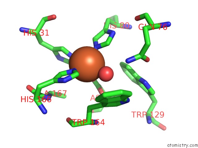
Mono view
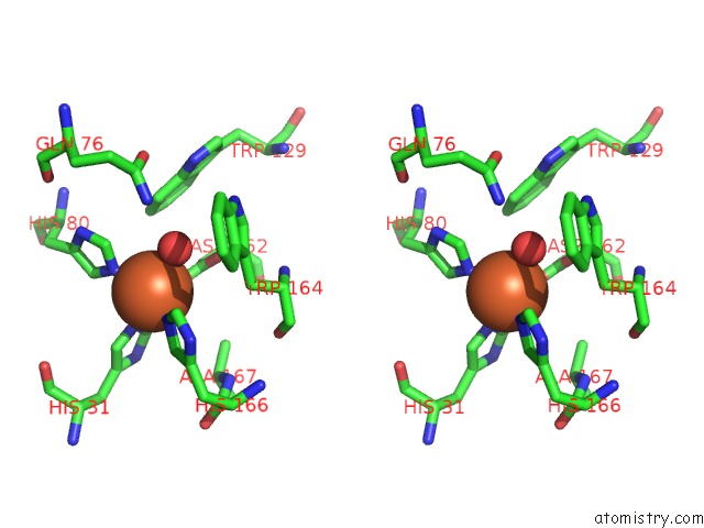
Stereo pair view
Mono view
Stereo pair view
A full contact list of Iron with other atoms in the Fe binding
site number 1 of Crystal Structure of Superoxide Dismutase From P. Marinus within 5.0Å range:
|
Iron binding site 2 out of 2 in 2cw2
Go back to
Iron binding site 2 out
of 2 in the Crystal Structure of Superoxide Dismutase From P. Marinus
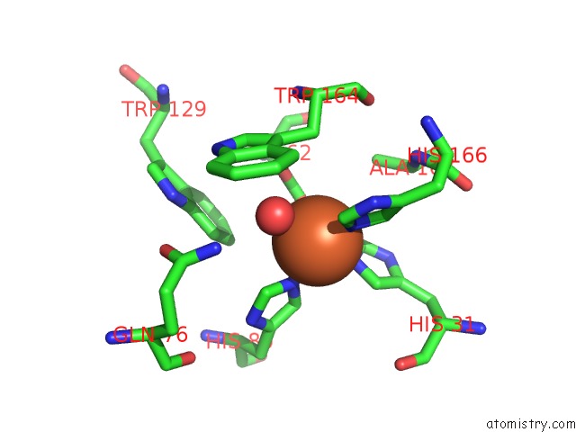
Mono view
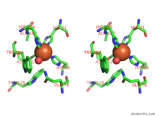
Stereo pair view
Mono view
Stereo pair view
A full contact list of Iron with other atoms in the Fe binding
site number 2 of Crystal Structure of Superoxide Dismutase From P. Marinus within 5.0Å range:
|
Reference:
O.A.Asojo,
E.J.Schott,
G.R.Vasta,
A.M.Silva.
Structures of PMSOD1 and PMSOD2, Two Superoxide Dismutases From the Protozoan Parasite Perkinsus Marinus Acta Crystallogr.,Sect.F V. 62 1072 2006.
ISSN: ESSN 1744-3091
PubMed: 17077482
DOI: 10.1107/S1744309106040425
Page generated: Thu Jul 17 00:30:42 2025
ISSN: ESSN 1744-3091
PubMed: 17077482
DOI: 10.1107/S1744309106040425
Last articles
Na in 4O52Na in 4O4W
Na in 4O4X
Na in 4O4V
Na in 4O48
Na in 4O47
Na in 4O1R
Na in 4O1Q
Na in 4O34
Na in 4O0I