Iron »
PDB 2ciz-2d3q »
2cxb »
Iron in PDB 2cxb: Crystallization and X-Ray Structure Determination of Cytochrome C2 From Rhodobacter Sphaeroides in Three Crystal Forms
Protein crystallography data
The structure of Crystallization and X-Ray Structure Determination of Cytochrome C2 From Rhodobacter Sphaeroides in Three Crystal Forms, PDB code: 2cxb
was solved by
H.L.Axelrod,
G.Feher,
J.P.Allen,
A.J.Chirino,
M.W.Day,
B.T.Hsu,
D.C.Rees,
with X-Ray Crystallography technique. A brief refinement statistics is given in the table below:
| Resolution Low / High (Å) | 6.00 / 1.95 |
| Space group | P 1 |
| Cell size a, b, c (Å), α, β, γ (°) | 45.310, 37.970, 37.600, 102.94, 72.44, 90.57 |
| R / Rfree (%) | n/a / n/a |
Iron Binding Sites:
The binding sites of Iron atom in the Crystallization and X-Ray Structure Determination of Cytochrome C2 From Rhodobacter Sphaeroides in Three Crystal Forms
(pdb code 2cxb). This binding sites where shown within
5.0 Angstroms radius around Iron atom.
In total 2 binding sites of Iron where determined in the Crystallization and X-Ray Structure Determination of Cytochrome C2 From Rhodobacter Sphaeroides in Three Crystal Forms, PDB code: 2cxb:
Jump to Iron binding site number: 1; 2;
In total 2 binding sites of Iron where determined in the Crystallization and X-Ray Structure Determination of Cytochrome C2 From Rhodobacter Sphaeroides in Three Crystal Forms, PDB code: 2cxb:
Jump to Iron binding site number: 1; 2;
Iron binding site 1 out of 2 in 2cxb
Go back to
Iron binding site 1 out
of 2 in the Crystallization and X-Ray Structure Determination of Cytochrome C2 From Rhodobacter Sphaeroides in Three Crystal Forms
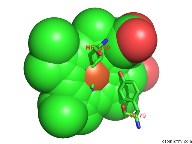
Mono view
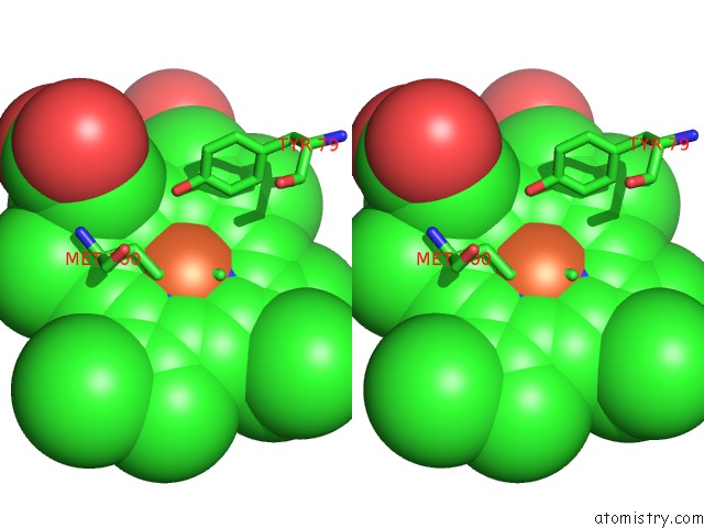
Stereo pair view
Mono view
Stereo pair view
A full contact list of Iron with other atoms in the Fe binding
site number 1 of Crystallization and X-Ray Structure Determination of Cytochrome C2 From Rhodobacter Sphaeroides in Three Crystal Forms within 5.0Å range:
|
Iron binding site 2 out of 2 in 2cxb
Go back to
Iron binding site 2 out
of 2 in the Crystallization and X-Ray Structure Determination of Cytochrome C2 From Rhodobacter Sphaeroides in Three Crystal Forms
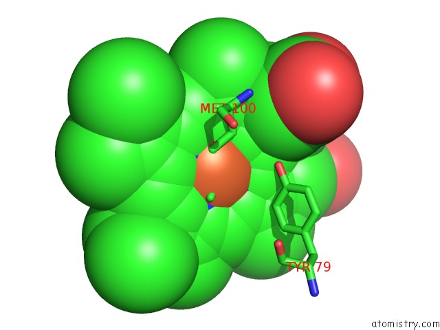
Mono view
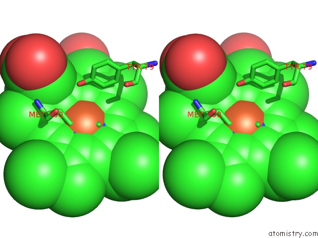
Stereo pair view
Mono view
Stereo pair view
A full contact list of Iron with other atoms in the Fe binding
site number 2 of Crystallization and X-Ray Structure Determination of Cytochrome C2 From Rhodobacter Sphaeroides in Three Crystal Forms within 5.0Å range:
|
Reference:
H.L.Axelrod,
G.Feher,
J.P.Allen,
A.J.Chirino,
M.W.Day,
B.T.Hsu,
D.C.Rees.
Crystallization and X-Ray Structure Determination of Cytochrome C2 From Rhodobacter Sphaeroides in Three Crystal Forms. Acta Crystallogr.,Sect.D V. 50 596 1994.
ISSN: ISSN 0907-4449
PubMed: 15299423
DOI: 10.1107/S0907444994001319
Page generated: Thu Jul 17 00:31:51 2025
ISSN: ISSN 0907-4449
PubMed: 15299423
DOI: 10.1107/S0907444994001319
Last articles
Na in 3MICNa in 3MI3
Na in 3MIE
Na in 3MGI
Na in 3ME4
Na in 3MDC
Na in 3MGA
Na in 3MEQ
Na in 3ME2
Na in 3MDA