Iron »
PDB 2e1s-2eus »
2eha »
Iron in PDB 2eha: Crystal Structure of Goat Lactoperoxidase Complexed with Formate Anion at 3.3 A Resolution
Enzymatic activity of Crystal Structure of Goat Lactoperoxidase Complexed with Formate Anion at 3.3 A Resolution
All present enzymatic activity of Crystal Structure of Goat Lactoperoxidase Complexed with Formate Anion at 3.3 A Resolution:
1.11.1.7;
1.11.1.7;
Protein crystallography data
The structure of Crystal Structure of Goat Lactoperoxidase Complexed with Formate Anion at 3.3 A Resolution, PDB code: 2eha
was solved by
A.K.Singh,
A.S.Ethayathulla,
N.Singh,
S.Sharma,
P.Kaur,
T.P.Singh,
with X-Ray Crystallography technique. A brief refinement statistics is given in the table below:
| Resolution Low / High (Å) | 20.00 / 3.30 |
| Space group | P 1 |
| Cell size a, b, c (Å), α, β, γ (°) | 57.991, 72.272, 83.652, 85.45, 84.04, 75.89 |
| R / Rfree (%) | 19.6 / 23.7 |
Other elements in 2eha:
The structure of Crystal Structure of Goat Lactoperoxidase Complexed with Formate Anion at 3.3 A Resolution also contains other interesting chemical elements:
| Calcium | (Ca) | 2 atoms |
Iron Binding Sites:
The binding sites of Iron atom in the Crystal Structure of Goat Lactoperoxidase Complexed with Formate Anion at 3.3 A Resolution
(pdb code 2eha). This binding sites where shown within
5.0 Angstroms radius around Iron atom.
In total 2 binding sites of Iron where determined in the Crystal Structure of Goat Lactoperoxidase Complexed with Formate Anion at 3.3 A Resolution, PDB code: 2eha:
Jump to Iron binding site number: 1; 2;
In total 2 binding sites of Iron where determined in the Crystal Structure of Goat Lactoperoxidase Complexed with Formate Anion at 3.3 A Resolution, PDB code: 2eha:
Jump to Iron binding site number: 1; 2;
Iron binding site 1 out of 2 in 2eha
Go back to
Iron binding site 1 out
of 2 in the Crystal Structure of Goat Lactoperoxidase Complexed with Formate Anion at 3.3 A Resolution
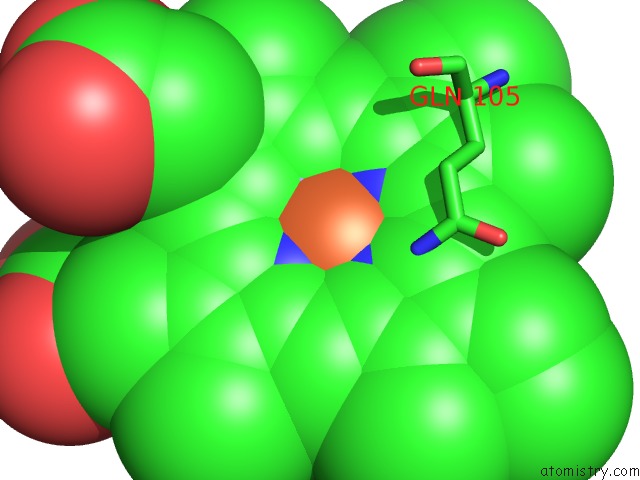
Mono view
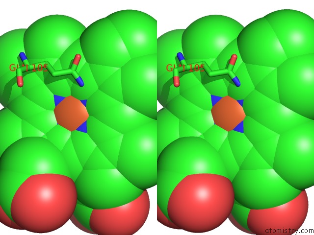
Stereo pair view
Mono view
Stereo pair view
A full contact list of Iron with other atoms in the Fe binding
site number 1 of Crystal Structure of Goat Lactoperoxidase Complexed with Formate Anion at 3.3 A Resolution within 5.0Å range:
|
Iron binding site 2 out of 2 in 2eha
Go back to
Iron binding site 2 out
of 2 in the Crystal Structure of Goat Lactoperoxidase Complexed with Formate Anion at 3.3 A Resolution
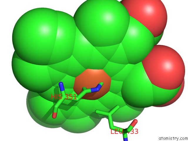
Mono view
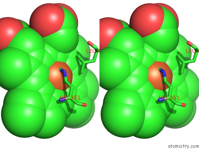
Stereo pair view
Mono view
Stereo pair view
A full contact list of Iron with other atoms in the Fe binding
site number 2 of Crystal Structure of Goat Lactoperoxidase Complexed with Formate Anion at 3.3 A Resolution within 5.0Å range:
|
Reference:
A.K.Singh,
A.S.Ethayathulla,
N.Singh,
S.Sharma,
A.Bhushan,
P.Kaur,
T.P.Singh.
Crystal Structure of Goat Lactoperoxidase Complexed with Formate Anion at 3.3 A Resolution To Be Published.
Page generated: Sat Aug 3 21:06:27 2024
Last articles
F in 9MIWF in 9IRP
F in 9IRM
F in 9MSQ
F in 9MEL
F in 9LRJ
F in 9M3P
F in 9LJ5
F in 9JVW
F in 9LJ1