Iron »
PDB 2eut-2fkz »
2fam »
Iron in PDB 2fam: X-Ray Crystal Structure of Ferric Aplysia Limacina Myoglobin in Different Liganded States
Protein crystallography data
The structure of X-Ray Crystal Structure of Ferric Aplysia Limacina Myoglobin in Different Liganded States, PDB code: 2fam
was solved by
E.Conti,
C.Moser,
M.Rizzi,
A.Mattevi,
C.Lionetti,
A.Coda,
P.Ascenzi,
M.Brunori,
M.Bolognesi,
with X-Ray Crystallography technique. A brief refinement statistics is given in the table below:
| Resolution Low / High (Å) | N/A / 2.00 |
| Space group | P 21 21 21 |
| Cell size a, b, c (Å), α, β, γ (°) | 52.980, 70.700, 32.500, 90.00, 90.00, 90.00 |
| R / Rfree (%) | n/a / n/a |
Iron Binding Sites:
The binding sites of Iron atom in the X-Ray Crystal Structure of Ferric Aplysia Limacina Myoglobin in Different Liganded States
(pdb code 2fam). This binding sites where shown within
5.0 Angstroms radius around Iron atom.
In total only one binding site of Iron was determined in the X-Ray Crystal Structure of Ferric Aplysia Limacina Myoglobin in Different Liganded States, PDB code: 2fam:
In total only one binding site of Iron was determined in the X-Ray Crystal Structure of Ferric Aplysia Limacina Myoglobin in Different Liganded States, PDB code: 2fam:
Iron binding site 1 out of 1 in 2fam
Go back to
Iron binding site 1 out
of 1 in the X-Ray Crystal Structure of Ferric Aplysia Limacina Myoglobin in Different Liganded States
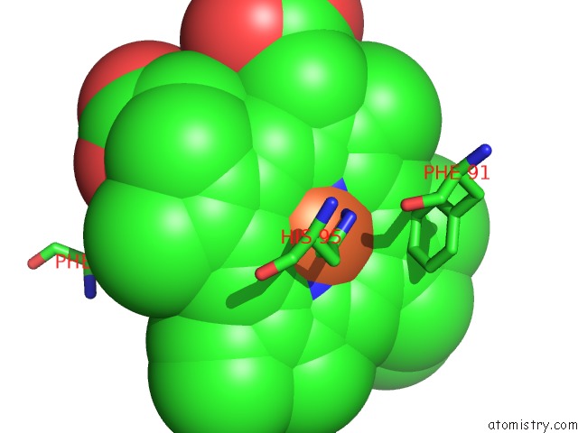
Mono view
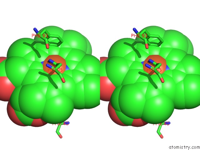
Stereo pair view
Mono view
Stereo pair view
A full contact list of Iron with other atoms in the Fe binding
site number 1 of X-Ray Crystal Structure of Ferric Aplysia Limacina Myoglobin in Different Liganded States within 5.0Å range:
|
Reference:
E.Conti,
C.Moser,
M.Rizzi,
A.Mattevi,
C.Lionetti,
A.Coda,
P.Ascenzi,
M.Brunori,
M.Bolognesi.
X-Ray Crystal Structure of Ferric Aplysia Limacina Myoglobin in Different Liganded States. J.Mol.Biol. V. 233 498 1993.
ISSN: ISSN 0022-2836
PubMed: 8411158
DOI: 10.1006/JMBI.1993.1527
Page generated: Thu Jul 17 00:58:05 2025
ISSN: ISSN 0022-2836
PubMed: 8411158
DOI: 10.1006/JMBI.1993.1527
Last articles
Zn in 1KNOZn in 1KMG
Zn in 1KL9
Zn in 1KLS
Zn in 1KLR
Zn in 1KL6
Zn in 1KLN
Zn in 1KK3
Zn in 1KKK
Zn in 1KK1