Iron »
PDB 2eut-2fkz »
2fdw »
Iron in PDB 2fdw: Crystal Structure of Human Microsomal P450 2A6 with the Inhibitor (5-(Pyridin-3-Yl)Furan-2-Yl)Methanamine Bound
Enzymatic activity of Crystal Structure of Human Microsomal P450 2A6 with the Inhibitor (5-(Pyridin-3-Yl)Furan-2-Yl)Methanamine Bound
All present enzymatic activity of Crystal Structure of Human Microsomal P450 2A6 with the Inhibitor (5-(Pyridin-3-Yl)Furan-2-Yl)Methanamine Bound:
1.14.14.1;
1.14.14.1;
Protein crystallography data
The structure of Crystal Structure of Human Microsomal P450 2A6 with the Inhibitor (5-(Pyridin-3-Yl)Furan-2-Yl)Methanamine Bound, PDB code: 2fdw
was solved by
J.K.Yano,
C.D.Stout,
E.F.Johnson,
with X-Ray Crystallography technique. A brief refinement statistics is given in the table below:
| Resolution Low / High (Å) | 34.59 / 2.05 |
| Space group | P 1 21 1 |
| Cell size a, b, c (Å), α, β, γ (°) | 69.746, 157.564, 103.789, 90.00, 91.88, 90.00 |
| R / Rfree (%) | 20.5 / 24.1 |
Iron Binding Sites:
The binding sites of Iron atom in the Crystal Structure of Human Microsomal P450 2A6 with the Inhibitor (5-(Pyridin-3-Yl)Furan-2-Yl)Methanamine Bound
(pdb code 2fdw). This binding sites where shown within
5.0 Angstroms radius around Iron atom.
In total 4 binding sites of Iron where determined in the Crystal Structure of Human Microsomal P450 2A6 with the Inhibitor (5-(Pyridin-3-Yl)Furan-2-Yl)Methanamine Bound, PDB code: 2fdw:
Jump to Iron binding site number: 1; 2; 3; 4;
In total 4 binding sites of Iron where determined in the Crystal Structure of Human Microsomal P450 2A6 with the Inhibitor (5-(Pyridin-3-Yl)Furan-2-Yl)Methanamine Bound, PDB code: 2fdw:
Jump to Iron binding site number: 1; 2; 3; 4;
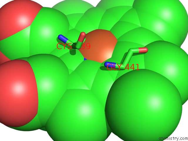
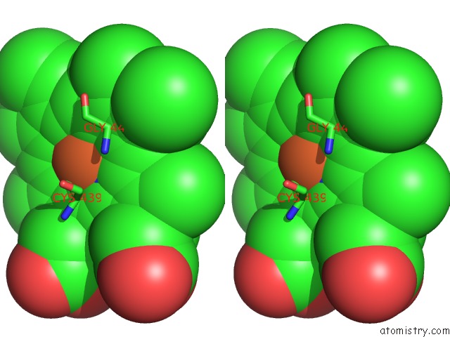
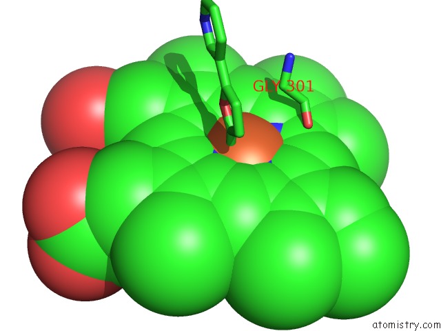
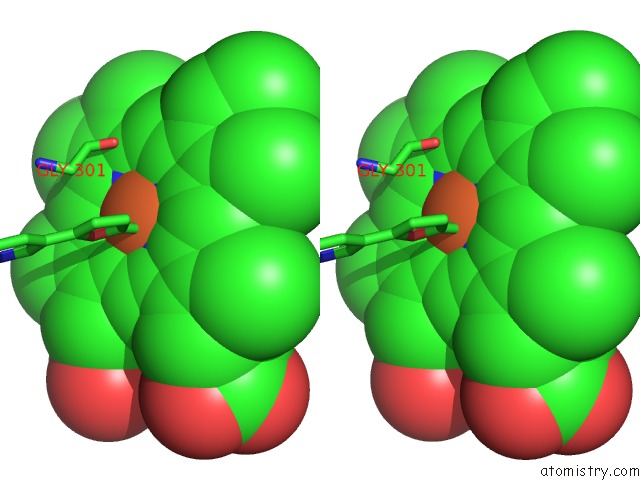
Iron binding site 1 out of 4 in 2fdw
Go back to
Iron binding site 1 out
of 4 in the Crystal Structure of Human Microsomal P450 2A6 with the Inhibitor (5-(Pyridin-3-Yl)Furan-2-Yl)Methanamine Bound
Mono view
Stereo pair view
Mono view
Stereo pair view
A full contact list of Iron with other atoms in the Fe binding
site number 1 of Crystal Structure of Human Microsomal P450 2A6 with the Inhibitor (5-(Pyridin-3-Yl)Furan-2-Yl)Methanamine Bound within 5.0Å range:
|
Iron binding site 2 out of 4 in 2fdw
Go back to
Iron binding site 2 out
of 4 in the Crystal Structure of Human Microsomal P450 2A6 with the Inhibitor (5-(Pyridin-3-Yl)Furan-2-Yl)Methanamine Bound
Mono view
Stereo pair view
Mono view
Stereo pair view
A full contact list of Iron with other atoms in the Fe binding
site number 2 of Crystal Structure of Human Microsomal P450 2A6 with the Inhibitor (5-(Pyridin-3-Yl)Furan-2-Yl)Methanamine Bound within 5.0Å range:
|
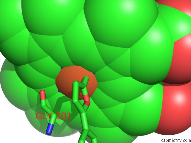
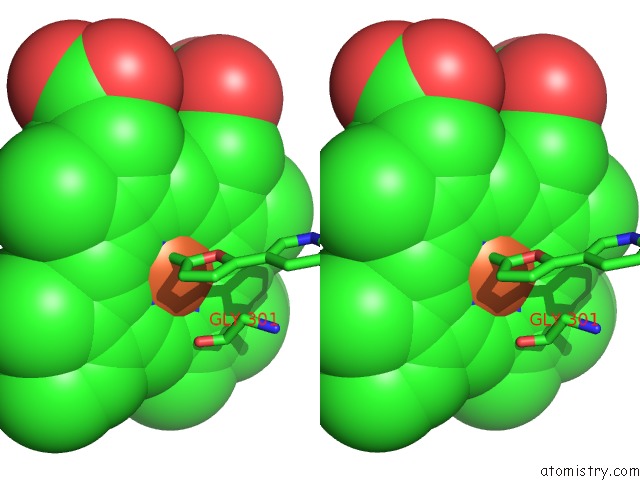
Iron binding site 3 out of 4 in 2fdw
Go back to
Iron binding site 3 out
of 4 in the Crystal Structure of Human Microsomal P450 2A6 with the Inhibitor (5-(Pyridin-3-Yl)Furan-2-Yl)Methanamine Bound
Mono view
Stereo pair view
Mono view
Stereo pair view
A full contact list of Iron with other atoms in the Fe binding
site number 3 of Crystal Structure of Human Microsomal P450 2A6 with the Inhibitor (5-(Pyridin-3-Yl)Furan-2-Yl)Methanamine Bound within 5.0Å range:
|
Iron binding site 4 out of 4 in 2fdw
Go back to
Iron binding site 4 out
of 4 in the Crystal Structure of Human Microsomal P450 2A6 with the Inhibitor (5-(Pyridin-3-Yl)Furan-2-Yl)Methanamine Bound
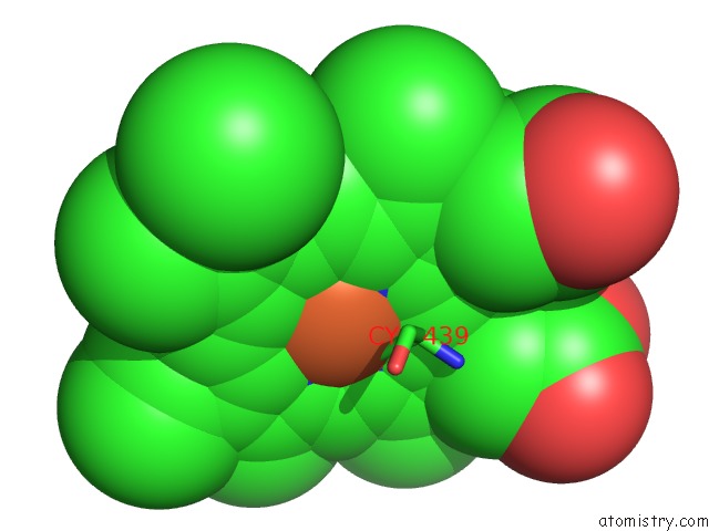
Mono view
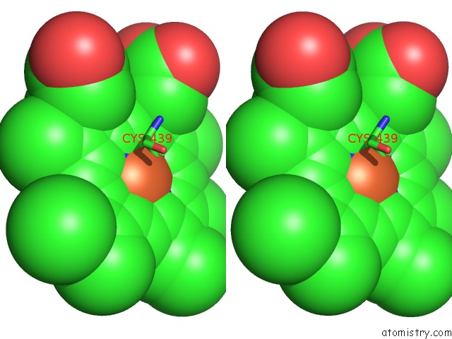
Stereo pair view
Mono view
Stereo pair view
A full contact list of Iron with other atoms in the Fe binding
site number 4 of Crystal Structure of Human Microsomal P450 2A6 with the Inhibitor (5-(Pyridin-3-Yl)Furan-2-Yl)Methanamine Bound within 5.0Å range:
|
Reference:
J.K.Yano,
T.T.Denton,
M.A.Cerny,
X.Zhang,
E.F.Johnson,
J.R.Cashman.
Synthetic Inhibitors of Cytochrome P-450 2A6: Inhibitory Activity, Difference Spectra, Mechanism of Inhibition, and Protein Cocrystallization. J.Med.Chem. V. 49 6987 2006.
ISSN: ISSN 0022-2623
PubMed: 17125252
DOI: 10.1021/JM060519R
Page generated: Thu Jul 17 01:04:26 2025
ISSN: ISSN 0022-2623
PubMed: 17125252
DOI: 10.1021/JM060519R
Last articles
Zn in 1JZQZn in 1JZS
Zn in 1JXP
Zn in 1JYB
Zn in 1JY8
Zn in 1JWH
Zn in 1JWQ
Zn in 1JV0
Zn in 1JWB
Zn in 1JW9