Iron »
PDB 2eut-2fkz »
2fjc »
Iron in PDB 2fjc: Crystal Structure of Antigen TPF1 From Treponema Pallidum
Protein crystallography data
The structure of Crystal Structure of Antigen TPF1 From Treponema Pallidum, PDB code: 2fjc
was solved by
A.Thumiger,
A.Polenghi,
E.Papinutto,
R.Battistutta,
C.Montecucco,
G.Zanotti,
with X-Ray Crystallography technique. A brief refinement statistics is given in the table below:
| Resolution Low / High (Å) | 59.37 / 2.50 |
| Space group | P 3 2 1 |
| Cell size a, b, c (Å), α, β, γ (°) | 184.921, 184.921, 154.883, 90.00, 90.00, 120.00 |
| R / Rfree (%) | 22.7 / 25.2 |
Iron Binding Sites:
Pages:
>>> Page 1 <<< Page 2, Binding sites: 11 - 16;Binding sites:
The binding sites of Iron atom in the Crystal Structure of Antigen TPF1 From Treponema Pallidum (pdb code 2fjc). This binding sites where shown within 5.0 Angstroms radius around Iron atom.In total 16 binding sites of Iron where determined in the Crystal Structure of Antigen TPF1 From Treponema Pallidum, PDB code: 2fjc:
Jump to Iron binding site number: 1; 2; 3; 4; 5; 6; 7; 8; 9; 10;
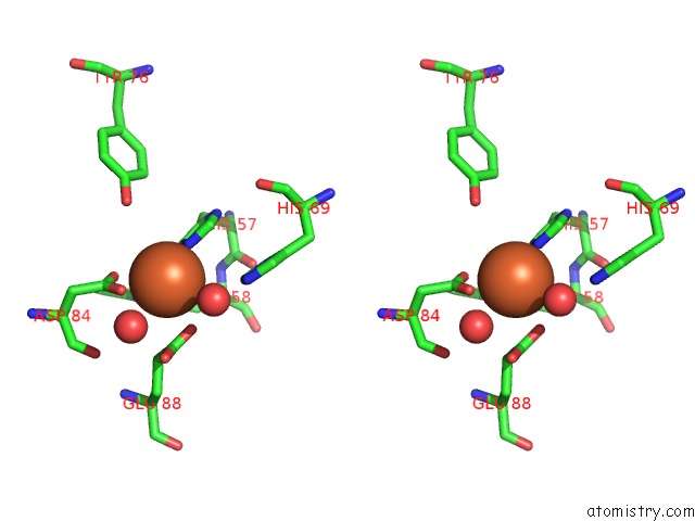
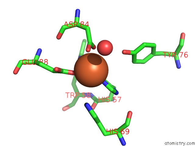
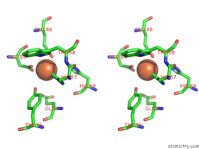
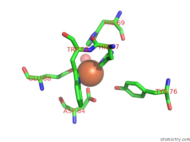
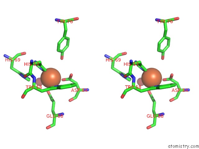
Iron binding site 1 out of 16 in 2fjc
Go back to
Iron binding site 1 out
of 16 in the Crystal Structure of Antigen TPF1 From Treponema Pallidum
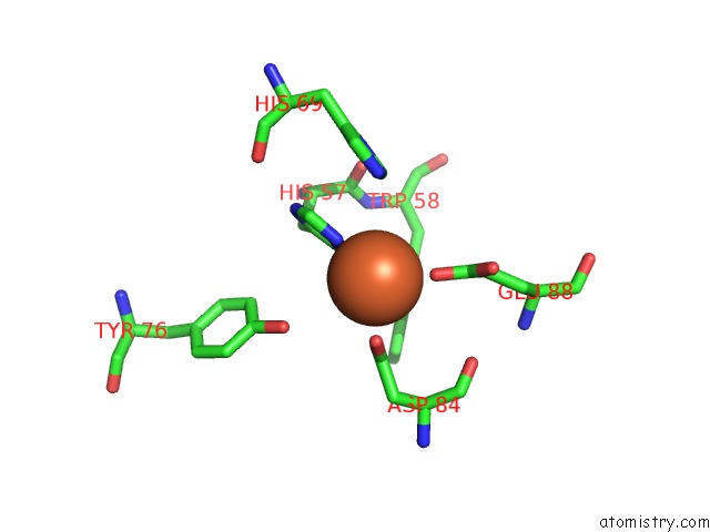
Mono view
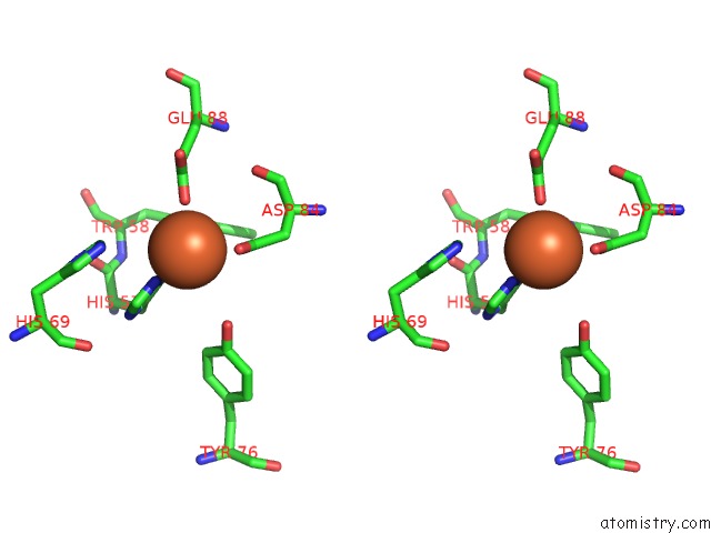
Stereo pair view
Mono view
Stereo pair view
A full contact list of Iron with other atoms in the Fe binding
site number 1 of Crystal Structure of Antigen TPF1 From Treponema Pallidum within 5.0Å range:
|
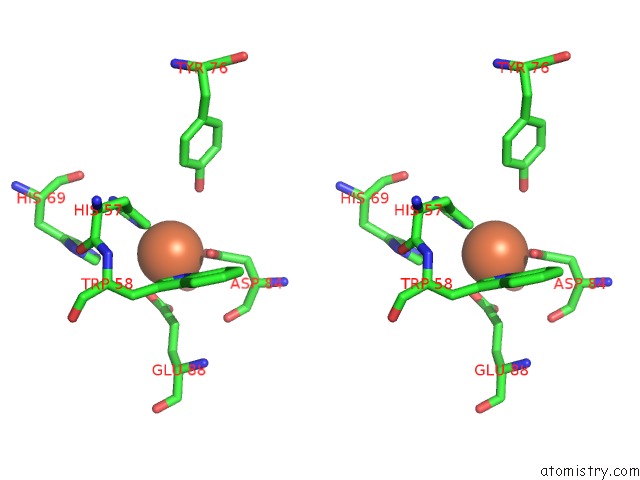
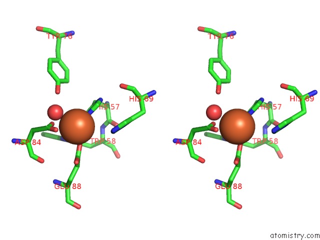
Iron binding site 2 out of 16 in 2fjc
Go back to
Iron binding site 2 out
of 16 in the Crystal Structure of Antigen TPF1 From Treponema Pallidum
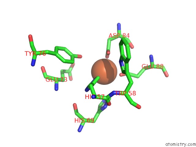
Mono view

Stereo pair view
Mono view
Stereo pair view
A full contact list of Iron with other atoms in the Fe binding
site number 2 of Crystal Structure of Antigen TPF1 From Treponema Pallidum within 5.0Å range:
|
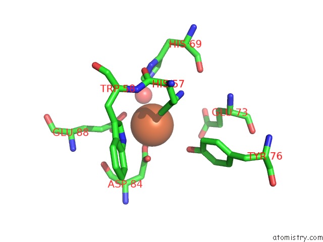
Iron binding site 3 out of 16 in 2fjc
Go back to
Iron binding site 3 out
of 16 in the Crystal Structure of Antigen TPF1 From Treponema Pallidum

Mono view
Stereo pair view
Mono view
Stereo pair view
A full contact list of Iron with other atoms in the Fe binding
site number 3 of Crystal Structure of Antigen TPF1 From Treponema Pallidum within 5.0Å range:
|
Iron binding site 4 out of 16 in 2fjc
Go back to
Iron binding site 4 out
of 16 in the Crystal Structure of Antigen TPF1 From Treponema Pallidum

Mono view
Stereo pair view
Mono view
Stereo pair view
A full contact list of Iron with other atoms in the Fe binding
site number 4 of Crystal Structure of Antigen TPF1 From Treponema Pallidum within 5.0Å range:
|
Iron binding site 5 out of 16 in 2fjc
Go back to
Iron binding site 5 out
of 16 in the Crystal Structure of Antigen TPF1 From Treponema Pallidum
Mono view
Stereo pair view
Mono view
Stereo pair view
A full contact list of Iron with other atoms in the Fe binding
site number 5 of Crystal Structure of Antigen TPF1 From Treponema Pallidum within 5.0Å range:
|
Iron binding site 6 out of 16 in 2fjc
Go back to
Iron binding site 6 out
of 16 in the Crystal Structure of Antigen TPF1 From Treponema Pallidum

Mono view

Stereo pair view
Mono view
Stereo pair view
A full contact list of Iron with other atoms in the Fe binding
site number 6 of Crystal Structure of Antigen TPF1 From Treponema Pallidum within 5.0Å range:
|
Iron binding site 7 out of 16 in 2fjc
Go back to
Iron binding site 7 out
of 16 in the Crystal Structure of Antigen TPF1 From Treponema Pallidum
Mono view

Stereo pair view
Mono view
Stereo pair view
A full contact list of Iron with other atoms in the Fe binding
site number 7 of Crystal Structure of Antigen TPF1 From Treponema Pallidum within 5.0Å range:
|
Iron binding site 8 out of 16 in 2fjc
Go back to
Iron binding site 8 out
of 16 in the Crystal Structure of Antigen TPF1 From Treponema Pallidum

Mono view
Stereo pair view
Mono view
Stereo pair view
A full contact list of Iron with other atoms in the Fe binding
site number 8 of Crystal Structure of Antigen TPF1 From Treponema Pallidum within 5.0Å range:
|
Iron binding site 9 out of 16 in 2fjc
Go back to
Iron binding site 9 out
of 16 in the Crystal Structure of Antigen TPF1 From Treponema Pallidum
Mono view
Stereo pair view
Mono view
Stereo pair view
A full contact list of Iron with other atoms in the Fe binding
site number 9 of Crystal Structure of Antigen TPF1 From Treponema Pallidum within 5.0Å range:
|
Iron binding site 10 out of 16 in 2fjc
Go back to
Iron binding site 10 out
of 16 in the Crystal Structure of Antigen TPF1 From Treponema Pallidum

Mono view

Stereo pair view
Mono view
Stereo pair view
A full contact list of Iron with other atoms in the Fe binding
site number 10 of Crystal Structure of Antigen TPF1 From Treponema Pallidum within 5.0Å range:
|
Reference:
A.Thumiger,
A.Polenghi,
E.Papinutto,
R.Battistutta,
C.Montecucco,
G.Zanotti.
Crystal Structure of Antigen TPF1 From Treponema Pallidum. Proteins V. 62 827 2006.
ISSN: ISSN 0887-3585
PubMed: 16345079
DOI: 10.1002/PROT.20828
Page generated: Thu Jul 17 01:07:28 2025
ISSN: ISSN 0887-3585
PubMed: 16345079
DOI: 10.1002/PROT.20828
Last articles
Zn in 1U0AZn in 1U0B
Zn in 1U05
Zn in 1TYM
Zn in 1TY2
Zn in 1TXR
Zn in 1TYL
Zn in 1TXL
Zn in 1TWG
Zn in 1TWC