Iron »
PDB 2fl0-2g6n »
2g0x »
Iron in PDB 2g0x: Photolyzed Co L29F Myoglobin: 316PS
Protein crystallography data
The structure of Photolyzed Co L29F Myoglobin: 316PS, PDB code: 2g0x
was solved by
R.Aranda,
E.J.Levin,
F.Schotte,
P.A.Anfinrud,
G.N.Phillips Jr.,
with X-Ray Crystallography technique. A brief refinement statistics is given in the table below:
| Resolution Low / High (Å) | 12.70 / 1.95 |
| Space group | P 6 |
| Cell size a, b, c (Å), α, β, γ (°) | 91.200, 91.200, 45.870, 90.00, 90.00, 120.00 |
| R / Rfree (%) | 5.5 / 6.3 |
Iron Binding Sites:
The binding sites of Iron atom in the Photolyzed Co L29F Myoglobin: 316PS
(pdb code 2g0x). This binding sites where shown within
5.0 Angstroms radius around Iron atom.
In total only one binding site of Iron was determined in the Photolyzed Co L29F Myoglobin: 316PS, PDB code: 2g0x:
In total only one binding site of Iron was determined in the Photolyzed Co L29F Myoglobin: 316PS, PDB code: 2g0x:
Iron binding site 1 out of 1 in 2g0x
Go back to
Iron binding site 1 out
of 1 in the Photolyzed Co L29F Myoglobin: 316PS
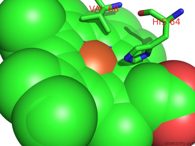
Mono view
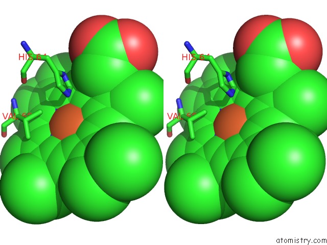
Stereo pair view
Mono view
Stereo pair view
A full contact list of Iron with other atoms in the Fe binding
site number 1 of Photolyzed Co L29F Myoglobin: 316PS within 5.0Å range:
|
Reference:
R.Aranda,
E.J.Levin,
F.Schotte,
P.A.Anfinrud,
G.N.Phillips Jr..
Time-Dependent Atomic Coordinates For the Dissociation of Carbon Monoxide From Myoglobin. Acta Crystallogr.,Sect.D V. 62 776 2006.
ISSN: ISSN 0907-4449
PubMed: 16790933
DOI: 10.1107/S0907444906017318
Page generated: Thu Jul 17 01:23:23 2025
ISSN: ISSN 0907-4449
PubMed: 16790933
DOI: 10.1107/S0907444906017318
Last articles
Zn in 1S3GZn in 1S39
Zn in 1S38
Zn in 1S30
Zn in 1S2Z
Zn in 1S1G
Zn in 1S0F
Zn in 1S0U
Zn in 1S0E
Zn in 1S0D