Iron »
PDB 2grz-2hk6 »
2hbg »
Iron in PDB 2hbg: Glycera Dibranchiata Hemoglobin. Structure and Refinement at 1.5 Angstroms Resolution
Protein crystallography data
The structure of Glycera Dibranchiata Hemoglobin. Structure and Refinement at 1.5 Angstroms Resolution, PDB code: 2hbg
was solved by
G.A.Arents,
W.E.Love,
with X-Ray Crystallography technique. A brief refinement statistics is given in the table below:
| Resolution Low / High (Å) | N/A / 1.50 |
| Space group | P 21 21 21 |
| Cell size a, b, c (Å), α, β, γ (°) | 42.750, 83.150, 38.660, 90.00, 90.00, 90.00 |
| R / Rfree (%) | n/a / n/a |
Iron Binding Sites:
The binding sites of Iron atom in the Glycera Dibranchiata Hemoglobin. Structure and Refinement at 1.5 Angstroms Resolution
(pdb code 2hbg). This binding sites where shown within
5.0 Angstroms radius around Iron atom.
In total only one binding site of Iron was determined in the Glycera Dibranchiata Hemoglobin. Structure and Refinement at 1.5 Angstroms Resolution, PDB code: 2hbg:
In total only one binding site of Iron was determined in the Glycera Dibranchiata Hemoglobin. Structure and Refinement at 1.5 Angstroms Resolution, PDB code: 2hbg:
Iron binding site 1 out of 1 in 2hbg
Go back to
Iron binding site 1 out
of 1 in the Glycera Dibranchiata Hemoglobin. Structure and Refinement at 1.5 Angstroms Resolution
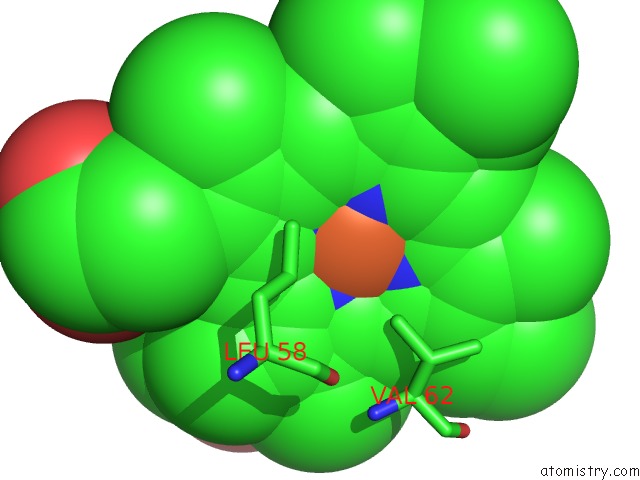
Mono view
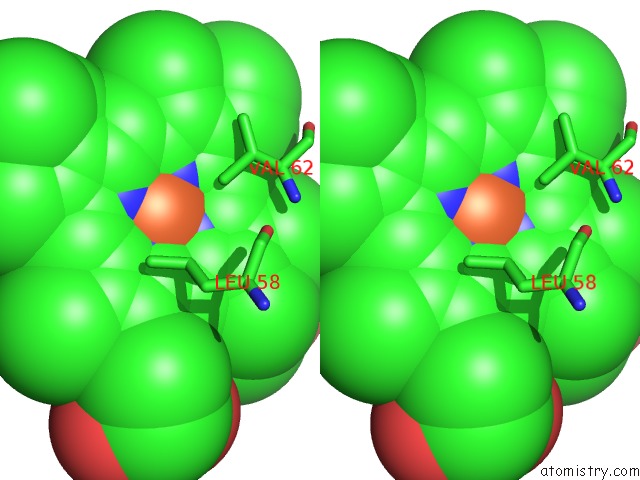
Stereo pair view
Mono view
Stereo pair view
A full contact list of Iron with other atoms in the Fe binding
site number 1 of Glycera Dibranchiata Hemoglobin. Structure and Refinement at 1.5 Angstroms Resolution within 5.0Å range:
|
Reference:
G.Arents,
W.E.Love.
Glycera Dibranchiata Hemoglobin. Structure and Refinement at 1.5 A Resolution. J.Mol.Biol. V. 210 149 1989.
ISSN: ISSN 0022-2836
PubMed: 2585515
DOI: 10.1016/0022-2836(89)90297-0
Page generated: Thu Jul 17 02:00:53 2025
ISSN: ISSN 0022-2836
PubMed: 2585515
DOI: 10.1016/0022-2836(89)90297-0
Last articles
Fe in 8CMOFe in 8CJY
Fe in 8CM7
Fe in 8CM6
Fe in 8CM4
Fe in 8CH9
Fe in 8CKS
Fe in 8CKN
Fe in 8CK9
Fe in 8CJO