Iron »
PDB 2grz-2hk6 »
2hhk »
Iron in PDB 2hhk: Reaction Centre From Rhodobacter Sphaeroides Strain R-26.1 Complexed with Dibrominated Phosphatidylglycerol
Protein crystallography data
The structure of Reaction Centre From Rhodobacter Sphaeroides Strain R-26.1 Complexed with Dibrominated Phosphatidylglycerol, PDB code: 2hhk
was solved by
A.W.Roszak,
A.T.Gardiner,
N.W.Isaacs,
R.J.Cogdell,
with X-Ray Crystallography technique. A brief refinement statistics is given in the table below:
| Resolution Low / High (Å) | 46.00 / 2.50 |
| Space group | P 31 2 1 |
| Cell size a, b, c (Å), α, β, γ (°) | 139.417, 139.417, 183.701, 90.00, 90.00, 120.00 |
| R / Rfree (%) | 17.2 / 19.7 |
Other elements in 2hhk:
The structure of Reaction Centre From Rhodobacter Sphaeroides Strain R-26.1 Complexed with Dibrominated Phosphatidylglycerol also contains other interesting chemical elements:
| Magnesium | (Mg) | 4 atoms |
| Potassium | (K) | 1 atom |
| Bromine | (Br) | 2 atoms |
| Chlorine | (Cl) | 1 atom |
Iron Binding Sites:
The binding sites of Iron atom in the Reaction Centre From Rhodobacter Sphaeroides Strain R-26.1 Complexed with Dibrominated Phosphatidylglycerol
(pdb code 2hhk). This binding sites where shown within
5.0 Angstroms radius around Iron atom.
In total only one binding site of Iron was determined in the Reaction Centre From Rhodobacter Sphaeroides Strain R-26.1 Complexed with Dibrominated Phosphatidylglycerol, PDB code: 2hhk:
In total only one binding site of Iron was determined in the Reaction Centre From Rhodobacter Sphaeroides Strain R-26.1 Complexed with Dibrominated Phosphatidylglycerol, PDB code: 2hhk:
Iron binding site 1 out of 1 in 2hhk
Go back to
Iron binding site 1 out
of 1 in the Reaction Centre From Rhodobacter Sphaeroides Strain R-26.1 Complexed with Dibrominated Phosphatidylglycerol
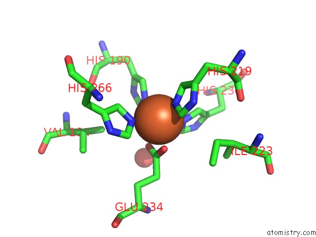
Mono view
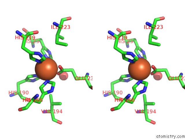
Stereo pair view
Mono view
Stereo pair view
|
|
A full contact list of Iron with other atoms in the Fe binding
site number 1 of Reaction Centre From Rhodobacter Sphaeroides Strain R-26.1 Complexed with Dibrominated Phosphatidylglycerol within 5.0Å range:
|
Reference:
A.W.Roszak,
A.T.Gardiner,
N.W.Isaacs,
R.J.Cogdell.
Brominated Lipids Identify Lipid Binding Sites on the Surface of the Reaction Center From Rhodobacter Sphaeroides. Biochemistry V. 46 2909 2007.
ISSN: ISSN 0006-2960
PubMed: 17315985
DOI: 10.1021/BI062154I
Page generated: Thu Jul 17 02:03:31 2025
ISSN: ISSN 0006-2960
PubMed: 17315985
DOI: 10.1021/BI062154I
Last articles
Zr in 1XC1Zr in 6Y7P
Zr in 6GNL
Zr in 6HYB
Zr in 4XYY
Zr in 5KHP
Zn in 9VXG
Zn in 9VWY
Zn in 9VCL
Zn in 9VKN