Iron »
PDB 2hkx-2ibn »
2ibn »
Iron in PDB 2ibn: Crystal Structure of Human Myo-Inositol Oxygenase (Miox)
Enzymatic activity of Crystal Structure of Human Myo-Inositol Oxygenase (Miox)
All present enzymatic activity of Crystal Structure of Human Myo-Inositol Oxygenase (Miox):
1.13.99.1;
1.13.99.1;
Protein crystallography data
The structure of Crystal Structure of Human Myo-Inositol Oxygenase (Miox), PDB code: 2ibn
was solved by
B.M.Hallberg,
R.D.Busam,
C.Arrowsmith,
H.Berglund,
R.Collins,
A.Edwards,
M.Ehn,
S.Flodin,
A.Flores,
S.Graslund,
M.Hammarstrom,
M.Hogbom,
L.Holmberg-Schiavone,
I.Johansson,
T.Karlberg,
T.Kotenyova,
P.Nilsson-Ehle,
P.Nordlund,
T.Nyman,
D.Ogg,
J.Sagemark,
P.Stenmark,
M.Sundstrom,
J.Uppenberg,
S.Van Den Berg,
J.Weigelt,
A.G.Thorsell,
C.Persson,
Structural Genomics Consortium (Sgc),
with X-Ray Crystallography technique. A brief refinement statistics is given in the table below:
| Resolution Low / High (Å) | 29.34 / 1.50 |
| Space group | C 1 2 1 |
| Cell size a, b, c (Å), α, β, γ (°) | 119.715, 55.861, 111.516, 90.00, 116.72, 90.00 |
| R / Rfree (%) | 20.6 / 24.8 |
Iron Binding Sites:
The binding sites of Iron atom in the Crystal Structure of Human Myo-Inositol Oxygenase (Miox)
(pdb code 2ibn). This binding sites where shown within
5.0 Angstroms radius around Iron atom.
In total 4 binding sites of Iron where determined in the Crystal Structure of Human Myo-Inositol Oxygenase (Miox), PDB code: 2ibn:
Jump to Iron binding site number: 1; 2; 3; 4;
In total 4 binding sites of Iron where determined in the Crystal Structure of Human Myo-Inositol Oxygenase (Miox), PDB code: 2ibn:
Jump to Iron binding site number: 1; 2; 3; 4;
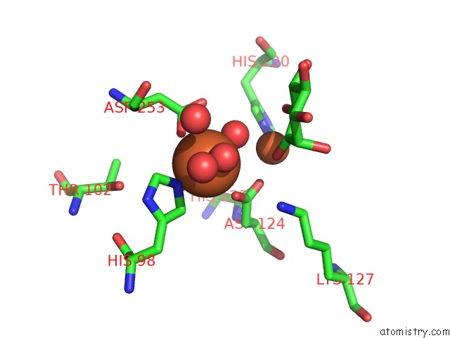
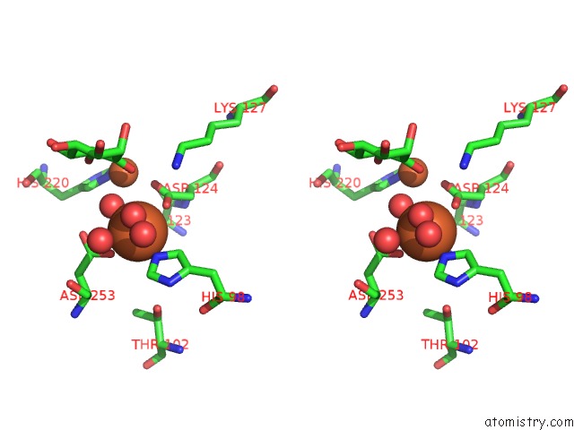
Iron binding site 1 out of 4 in 2ibn
Go back to
Iron binding site 1 out
of 4 in the Crystal Structure of Human Myo-Inositol Oxygenase (Miox)
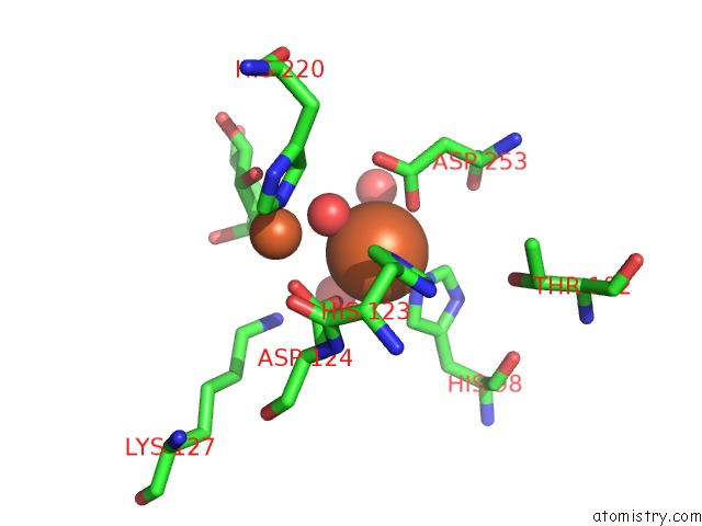
Mono view
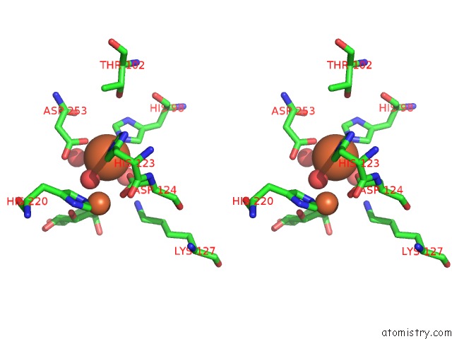
Stereo pair view
Mono view
Stereo pair view
A full contact list of Iron with other atoms in the Fe binding
site number 1 of Crystal Structure of Human Myo-Inositol Oxygenase (Miox) within 5.0Å range:
|
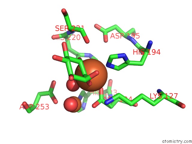
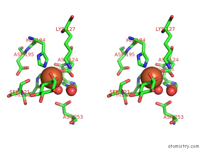
Iron binding site 2 out of 4 in 2ibn
Go back to
Iron binding site 2 out
of 4 in the Crystal Structure of Human Myo-Inositol Oxygenase (Miox)
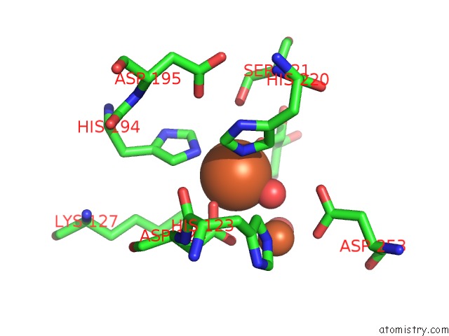
Mono view
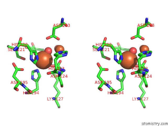
Stereo pair view
Mono view
Stereo pair view
A full contact list of Iron with other atoms in the Fe binding
site number 2 of Crystal Structure of Human Myo-Inositol Oxygenase (Miox) within 5.0Å range:
|
Iron binding site 3 out of 4 in 2ibn
Go back to
Iron binding site 3 out
of 4 in the Crystal Structure of Human Myo-Inositol Oxygenase (Miox)
Mono view
Stereo pair view
Mono view
Stereo pair view
A full contact list of Iron with other atoms in the Fe binding
site number 3 of Crystal Structure of Human Myo-Inositol Oxygenase (Miox) within 5.0Å range:
|
Iron binding site 4 out of 4 in 2ibn
Go back to
Iron binding site 4 out
of 4 in the Crystal Structure of Human Myo-Inositol Oxygenase (Miox)
Mono view
Stereo pair view
Mono view
Stereo pair view
A full contact list of Iron with other atoms in the Fe binding
site number 4 of Crystal Structure of Human Myo-Inositol Oxygenase (Miox) within 5.0Å range:
|
Reference:
A.G.Thorsell,
C.Persson,
N.Voevodskaya,
R.D.Busam,
M.Hammarstrom,
S.Graslund,
A.Graslund,
B.M.Hallberg.
Structural and Biophysical Characterization of Human Myo-Inositol Oxygenase J.Biol.Chem. V. 283 15209 2008.
ISSN: ISSN 0021-9258
PubMed: 18364358
DOI: 10.1074/JBC.M800348200
Page generated: Thu Jul 17 02:14:28 2025
ISSN: ISSN 0021-9258
PubMed: 18364358
DOI: 10.1074/JBC.M800348200
Last articles
W in 9FPPW in 8PRM
W in 9QM1
W in 9QM0
W in 9OJ3
W in 9MQX
W in 9FP4
W in 9BEO
W in 9BEM
W in 8P2U