Iron »
PDB 2j2m-2ksu »
2j9b »
Iron in PDB 2j9b: The Crystal Structure of Cytochrome C' From Rubrivivax Gelatinosus at 1.5 A Resolution and pH 6.3
Protein crystallography data
The structure of The Crystal Structure of Cytochrome C' From Rubrivivax Gelatinosus at 1.5 A Resolution and pH 6.3, PDB code: 2j9b
was solved by
S.Benini,
S.Ciurli,
W.R.Rypniewski,
K.S.Wilson,
with X-Ray Crystallography technique. A brief refinement statistics is given in the table below:
| Resolution Low / High (Å) | 19.92 / 1.50 |
| Space group | P 31 2 1 |
| Cell size a, b, c (Å), α, β, γ (°) | 69.558, 69.558, 123.374, 90.00, 90.00, 120.00 |
| R / Rfree (%) | 15.3 / 17.7 |
Iron Binding Sites:
The binding sites of Iron atom in the The Crystal Structure of Cytochrome C' From Rubrivivax Gelatinosus at 1.5 A Resolution and pH 6.3
(pdb code 2j9b). This binding sites where shown within
5.0 Angstroms radius around Iron atom.
In total 2 binding sites of Iron where determined in the The Crystal Structure of Cytochrome C' From Rubrivivax Gelatinosus at 1.5 A Resolution and pH 6.3, PDB code: 2j9b:
Jump to Iron binding site number: 1; 2;
In total 2 binding sites of Iron where determined in the The Crystal Structure of Cytochrome C' From Rubrivivax Gelatinosus at 1.5 A Resolution and pH 6.3, PDB code: 2j9b:
Jump to Iron binding site number: 1; 2;
Iron binding site 1 out of 2 in 2j9b
Go back to
Iron binding site 1 out
of 2 in the The Crystal Structure of Cytochrome C' From Rubrivivax Gelatinosus at 1.5 A Resolution and pH 6.3
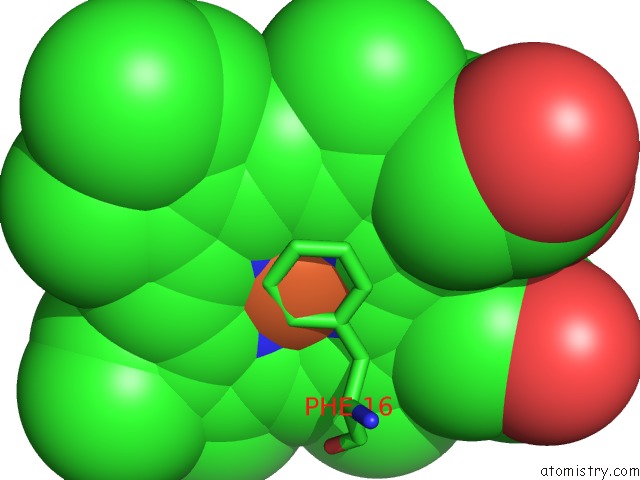
Mono view
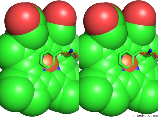
Stereo pair view
Mono view
Stereo pair view
A full contact list of Iron with other atoms in the Fe binding
site number 1 of The Crystal Structure of Cytochrome C' From Rubrivivax Gelatinosus at 1.5 A Resolution and pH 6.3 within 5.0Å range:
|
Iron binding site 2 out of 2 in 2j9b
Go back to
Iron binding site 2 out
of 2 in the The Crystal Structure of Cytochrome C' From Rubrivivax Gelatinosus at 1.5 A Resolution and pH 6.3
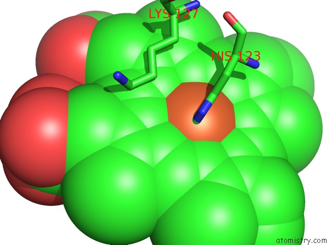
Mono view
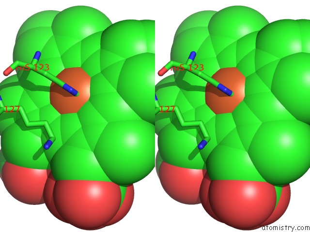
Stereo pair view
Mono view
Stereo pair view
A full contact list of Iron with other atoms in the Fe binding
site number 2 of The Crystal Structure of Cytochrome C' From Rubrivivax Gelatinosus at 1.5 A Resolution and pH 6.3 within 5.0Å range:
|
Reference:
S.Benini,
W.R.Rypniewski,
K.S.Wilson,
S.Ciurli.
High Resolution Crystal Structure of Rubrivivax Gelatinosus Cytochrome C'. J. Inorg. Biochem. V. 102 1322 2008.
ISSN: ISSN 0162-0134
PubMed: 18295896
DOI: 10.1016/J.JINORGBIO.2008.01.017
Page generated: Thu Jul 17 02:27:07 2025
ISSN: ISSN 0162-0134
PubMed: 18295896
DOI: 10.1016/J.JINORGBIO.2008.01.017
Last articles
K in 7QQRK in 7QQS
K in 7QQQ
K in 7QQP
K in 7QQO
K in 7QK5
K in 7QIX
K in 7QNO
K in 7QIY
K in 7Q3X