Iron »
PDB 2l4d-2mta »
2lhb »
Iron in PDB 2lhb: Refinement of A Molecular Model For Lamprey Hemoglobin From Petromyzon Marinus
Protein crystallography data
The structure of Refinement of A Molecular Model For Lamprey Hemoglobin From Petromyzon Marinus, PDB code: 2lhb
was solved by
R.B.Honzatko,
W.A.Hendrickson,
W.E.Love,
with X-Ray Crystallography technique. A brief refinement statistics is given in the table below:
| Resolution Low / High (Å) | N/A / 2.00 |
| Space group | P 21 21 21 |
| Cell size a, b, c (Å), α, β, γ (°) | 44.570, 96.620, 31.340, 90.00, 90.00, 90.00 |
| R / Rfree (%) | 14.2 / n/a |
Iron Binding Sites:
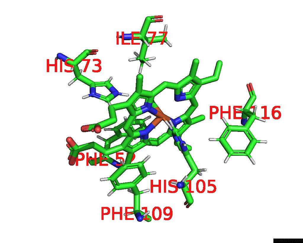
The binding sites of Iron atom in the Refinement of A Molecular Model For Lamprey Hemoglobin From Petromyzon Marinus
(pdb code 2lhb). This binding sites where shown within
5.0 Angstroms radius around Iron atom.
In total only one binding site of Iron was determined in the Refinement of A Molecular Model For Lamprey Hemoglobin From Petromyzon Marinus, PDB code: 2lhb:
In total only one binding site of Iron was determined in the Refinement of A Molecular Model For Lamprey Hemoglobin From Petromyzon Marinus, PDB code: 2lhb:
Iron binding site 1 out of 1 in 2lhb
Go back to
Iron binding site 1 out
of 1 in the Refinement of A Molecular Model For Lamprey Hemoglobin From Petromyzon Marinus
Mono view
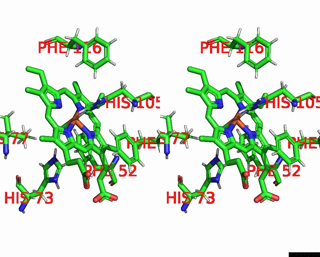
Stereo pair view
Mono view
Stereo pair view
A full contact list of Iron with other atoms in the Fe binding
site number 1 of Refinement of A Molecular Model For Lamprey Hemoglobin From Petromyzon Marinus within 5.0Å range:
|
Reference:
R.B.Honzatko,
W.A.Hendrickson,
W.E.Love.
Refinement of A Molecular Model For Lamprey Hemoglobin From Petromyzon Marinus. J.Mol.Biol. V. 184 147 1985.
ISSN: ISSN 0022-2836
PubMed: 4032476
DOI: 10.1016/0022-2836(85)90049-X
Page generated: Thu Jul 17 02:51:27 2025
ISSN: ISSN 0022-2836
PubMed: 4032476
DOI: 10.1016/0022-2836(85)90049-X
Last articles
Zn in 9QM9Zn in 9S44
Zn in 9OFE
Zn in 9OFC
Zn in 9OFD
Zn in 9OF1
Zn in 9OFB
Zn in 9N0J
Zn in 9M5X
Zn in 9LGI