Iron »
PDB 2l4d-2mta »
2mhb »
Iron in PDB 2mhb: The Structure of Horse Methaemoglobin at 2.0 Angstroms Resolution
Protein crystallography data
The structure of The Structure of Horse Methaemoglobin at 2.0 Angstroms Resolution, PDB code: 2mhb
was solved by
R.C.Ladner,
E.G.Heidner,
M.F.Perutz,
with X-Ray Crystallography technique. A brief refinement statistics is given in the table below:
| Resolution Low / High (Å) | N/A / 2.00 |
| Space group | C 1 2 1 |
| Cell size a, b, c (Å), α, β, γ (°) | 108.240, 63.130, 54.540, 90.00, 110.85, 90.00 |
| R / Rfree (%) | 23 / n/a |
Iron Binding Sites:
The binding sites of Iron atom in the The Structure of Horse Methaemoglobin at 2.0 Angstroms Resolution
(pdb code 2mhb). This binding sites where shown within
5.0 Angstroms radius around Iron atom.
In total 2 binding sites of Iron where determined in the The Structure of Horse Methaemoglobin at 2.0 Angstroms Resolution, PDB code: 2mhb:
Jump to Iron binding site number: 1; 2;
In total 2 binding sites of Iron where determined in the The Structure of Horse Methaemoglobin at 2.0 Angstroms Resolution, PDB code: 2mhb:
Jump to Iron binding site number: 1; 2;
Iron binding site 1 out of 2 in 2mhb
Go back to
Iron binding site 1 out
of 2 in the The Structure of Horse Methaemoglobin at 2.0 Angstroms Resolution
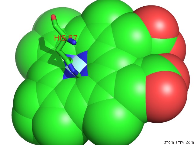
Mono view
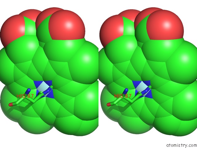
Stereo pair view
Mono view
Stereo pair view
A full contact list of Iron with other atoms in the Fe binding
site number 1 of The Structure of Horse Methaemoglobin at 2.0 Angstroms Resolution within 5.0Å range:
|
Iron binding site 2 out of 2 in 2mhb
Go back to
Iron binding site 2 out
of 2 in the The Structure of Horse Methaemoglobin at 2.0 Angstroms Resolution
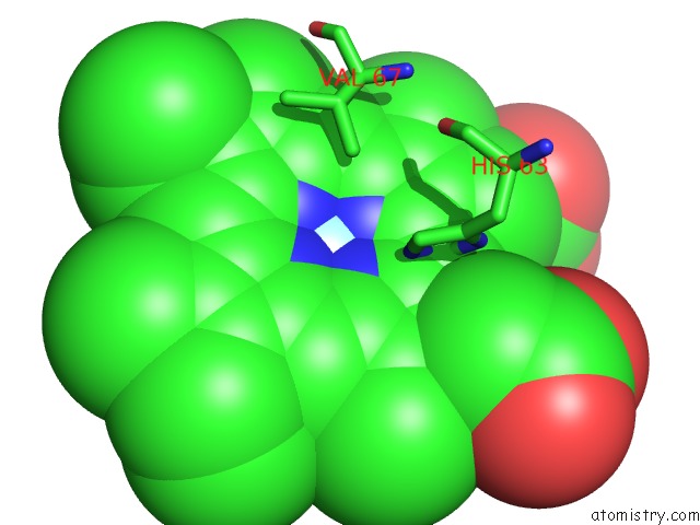
Mono view
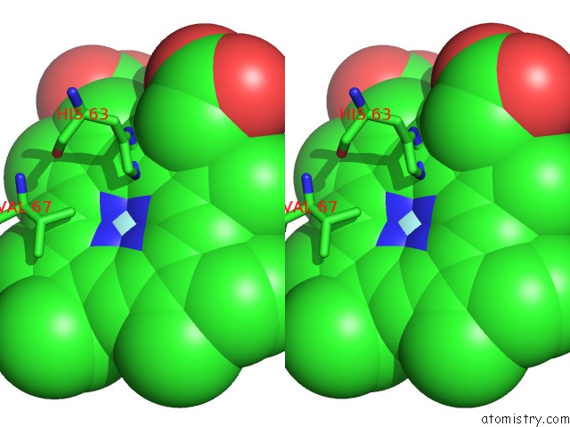
Stereo pair view
Mono view
Stereo pair view
A full contact list of Iron with other atoms in the Fe binding
site number 2 of The Structure of Horse Methaemoglobin at 2.0 Angstroms Resolution within 5.0Å range:
|
Reference:
R.C.Ladner,
E.J.Heidner,
M.F.Perutz.
The Structure of Horse Methaemoglobin at 2-0 A Resolution. J.Mol.Biol. V. 114 385 1977.
ISSN: ISSN 0022-2836
PubMed: 561852
DOI: 10.1016/0022-2836(77)90256-X
Page generated: Thu Jul 17 02:54:42 2025
ISSN: ISSN 0022-2836
PubMed: 561852
DOI: 10.1016/0022-2836(77)90256-X
Last articles
Fe in 2YXOFe in 2YRS
Fe in 2YXC
Fe in 2YNM
Fe in 2YVJ
Fe in 2YP1
Fe in 2YU2
Fe in 2YU1
Fe in 2YQB
Fe in 2YOO