Iron »
PDB 2mya-2nwb »
2nuk »
Iron in PDB 2nuk: Soluble Domain of the Rieske Iron-Sulfur Protein From Rhodobacter Sphaeroides
Enzymatic activity of Soluble Domain of the Rieske Iron-Sulfur Protein From Rhodobacter Sphaeroides
All present enzymatic activity of Soluble Domain of the Rieske Iron-Sulfur Protein From Rhodobacter Sphaeroides:
1.10.2.2;
1.10.2.2;
Protein crystallography data
The structure of Soluble Domain of the Rieske Iron-Sulfur Protein From Rhodobacter Sphaeroides, PDB code: 2nuk
was solved by
D.Kolling,
J.Brunzelle,
S.Lhee,
A.R.Crofts,
S.K.Nair,
with X-Ray Crystallography technique. A brief refinement statistics is given in the table below:
| Resolution Low / High (Å) | 11.67 / 1.20 |
| Space group | I 4 |
| Cell size a, b, c (Å), α, β, γ (°) | 70.568, 70.568, 54.769, 90.00, 90.00, 90.00 |
| R / Rfree (%) | 11.8 / 13.6 |
Iron Binding Sites:
The binding sites of Iron atom in the Soluble Domain of the Rieske Iron-Sulfur Protein From Rhodobacter Sphaeroides
(pdb code 2nuk). This binding sites where shown within
5.0 Angstroms radius around Iron atom.
In total 2 binding sites of Iron where determined in the Soluble Domain of the Rieske Iron-Sulfur Protein From Rhodobacter Sphaeroides, PDB code: 2nuk:
Jump to Iron binding site number: 1; 2;
In total 2 binding sites of Iron where determined in the Soluble Domain of the Rieske Iron-Sulfur Protein From Rhodobacter Sphaeroides, PDB code: 2nuk:
Jump to Iron binding site number: 1; 2;
Iron binding site 1 out of 2 in 2nuk
Go back to
Iron binding site 1 out
of 2 in the Soluble Domain of the Rieske Iron-Sulfur Protein From Rhodobacter Sphaeroides
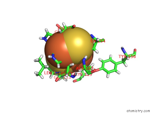
Mono view
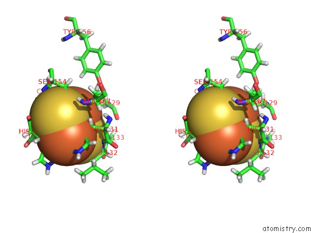
Stereo pair view
Mono view
Stereo pair view
A full contact list of Iron with other atoms in the Fe binding
site number 1 of Soluble Domain of the Rieske Iron-Sulfur Protein From Rhodobacter Sphaeroides within 5.0Å range:
|
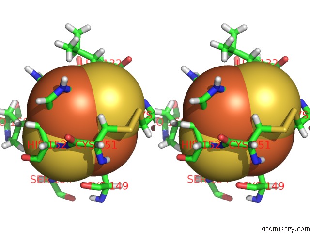
Iron binding site 2 out of 2 in 2nuk
Go back to
Iron binding site 2 out
of 2 in the Soluble Domain of the Rieske Iron-Sulfur Protein From Rhodobacter Sphaeroides
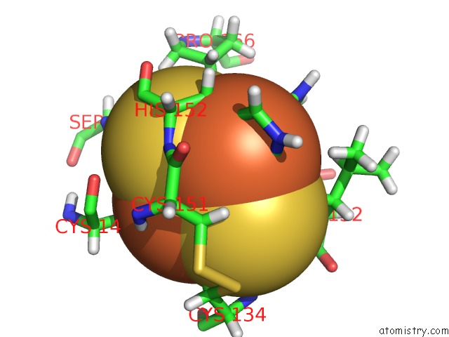
Mono view
Stereo pair view
Mono view
Stereo pair view
A full contact list of Iron with other atoms in the Fe binding
site number 2 of Soluble Domain of the Rieske Iron-Sulfur Protein From Rhodobacter Sphaeroides within 5.0Å range:
|
Reference:
D.J.Kolling,
J.S.Brunzelle,
S.Lhee,
A.R.Crofts,
S.K.Nair.
Atomic Resolution Structures of Rieske Iron-Sulfur Protein: Role of Hydrogen Bonds in Tuning the Redox Potential of Iron-Sulfur Clusters. Structure V. 15 29 2007.
ISSN: ISSN 0969-2126
PubMed: 17223530
DOI: 10.1016/J.STR.2006.11.012
Page generated: Thu Jul 17 03:03:49 2025
ISSN: ISSN 0969-2126
PubMed: 17223530
DOI: 10.1016/J.STR.2006.11.012
Last articles
Na in 6TBINa in 6TCJ
Na in 6TBT
Na in 6TBN
Na in 6TBF
Na in 6TBH
Na in 6TBG
Na in 6TB7
Na in 6TB1
Na in 6T99