Iron »
PDB 2nwf-2oof »
2oc5 »
Iron in PDB 2oc5: Crystal Structure of A Ferritin-Like Protein (PMT1231) From Prochlorococcus Marinus Str. Mit 9313 at 1.68 A Resolution
Protein crystallography data
The structure of Crystal Structure of A Ferritin-Like Protein (PMT1231) From Prochlorococcus Marinus Str. Mit 9313 at 1.68 A Resolution, PDB code: 2oc5
was solved by
Joint Center For Structural Genomics (Jcsg),
with X-Ray Crystallography technique. A brief refinement statistics is given in the table below:
| Resolution Low / High (Å) | 29.24 / 1.68 |
| Space group | P 43 21 2 |
| Cell size a, b, c (Å), α, β, γ (°) | 77.350, 77.350, 116.890, 90.00, 90.00, 90.00 |
| R / Rfree (%) | 16.4 / 20 |
Iron Binding Sites:
The binding sites of Iron atom in the Crystal Structure of A Ferritin-Like Protein (PMT1231) From Prochlorococcus Marinus Str. Mit 9313 at 1.68 A Resolution
(pdb code 2oc5). This binding sites where shown within
5.0 Angstroms radius around Iron atom.
In total 2 binding sites of Iron where determined in the Crystal Structure of A Ferritin-Like Protein (PMT1231) From Prochlorococcus Marinus Str. Mit 9313 at 1.68 A Resolution, PDB code: 2oc5:
Jump to Iron binding site number: 1; 2;
In total 2 binding sites of Iron where determined in the Crystal Structure of A Ferritin-Like Protein (PMT1231) From Prochlorococcus Marinus Str. Mit 9313 at 1.68 A Resolution, PDB code: 2oc5:
Jump to Iron binding site number: 1; 2;
Iron binding site 1 out of 2 in 2oc5
Go back to
Iron binding site 1 out
of 2 in the Crystal Structure of A Ferritin-Like Protein (PMT1231) From Prochlorococcus Marinus Str. Mit 9313 at 1.68 A Resolution
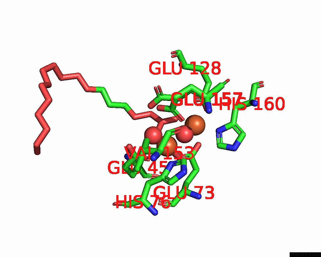
Mono view
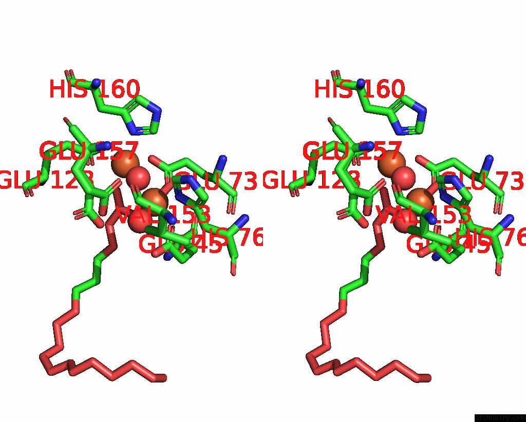
Stereo pair view
Mono view
Stereo pair view
A full contact list of Iron with other atoms in the Fe binding
site number 1 of Crystal Structure of A Ferritin-Like Protein (PMT1231) From Prochlorococcus Marinus Str. Mit 9313 at 1.68 A Resolution within 5.0Å range:
|
Iron binding site 2 out of 2 in 2oc5
Go back to
Iron binding site 2 out
of 2 in the Crystal Structure of A Ferritin-Like Protein (PMT1231) From Prochlorococcus Marinus Str. Mit 9313 at 1.68 A Resolution
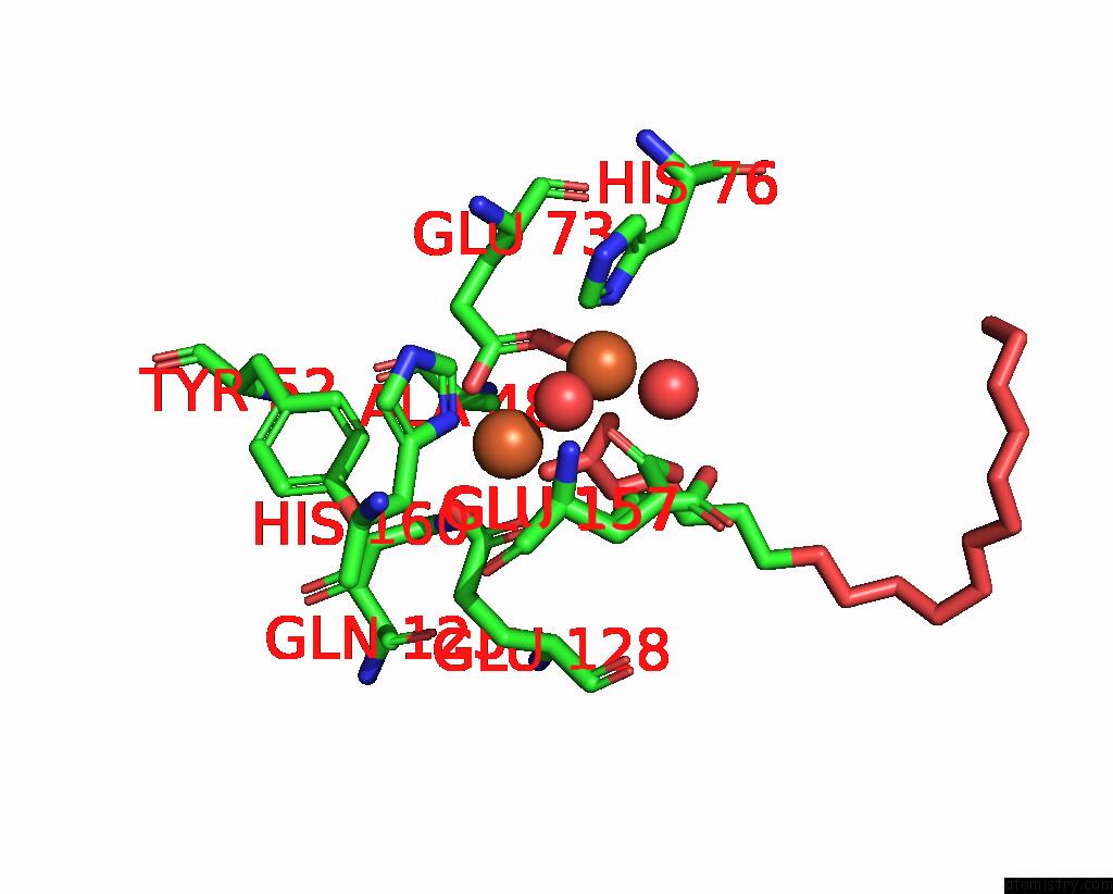
Mono view

Stereo pair view
Mono view
Stereo pair view
A full contact list of Iron with other atoms in the Fe binding
site number 2 of Crystal Structure of A Ferritin-Like Protein (PMT1231) From Prochlorococcus Marinus Str. Mit 9313 at 1.68 A Resolution within 5.0Å range:
|
Reference:
Joint Center For Structural Genomics (Jcsg),
Joint Center For Structural Genomics (Jcsg).
N/A N/A.
Page generated: Thu Jul 17 03:10:27 2025
Last articles
Mg in 382DMg in 370D
Mg in 360D
Mg in 336D
Mg in 355D
Mg in 308D
Mg in 335D
Mg in 310D
Mg in 314D
Mg in 313D