Iron »
PDB 2nwf-2oof »
2ofr »
Iron in PDB 2ofr: 1.00 A Crystal Structure of V36A/D129A/L130A Mutant of Nitrophorin 4 From Rhodnius Prolixus Complexed with Nitric Oxide at pH 5.6
Protein crystallography data
The structure of 1.00 A Crystal Structure of V36A/D129A/L130A Mutant of Nitrophorin 4 From Rhodnius Prolixus Complexed with Nitric Oxide at pH 5.6, PDB code: 2ofr
was solved by
A.M.Amoia,
with X-Ray Crystallography technique. A brief refinement statistics is given in the table below:
| Resolution Low / High (Å) | 9.47 / 1.00 |
| Space group | C 1 2 1 |
| Cell size a, b, c (Å), α, β, γ (°) | 70.191, 42.637, 52.975, 90.00, 94.12, 90.00 |
| R / Rfree (%) | 12.6 / 14.8 |
Iron Binding Sites:
The binding sites of Iron atom in the 1.00 A Crystal Structure of V36A/D129A/L130A Mutant of Nitrophorin 4 From Rhodnius Prolixus Complexed with Nitric Oxide at pH 5.6
(pdb code 2ofr). This binding sites where shown within
5.0 Angstroms radius around Iron atom.
In total only one binding site of Iron was determined in the 1.00 A Crystal Structure of V36A/D129A/L130A Mutant of Nitrophorin 4 From Rhodnius Prolixus Complexed with Nitric Oxide at pH 5.6, PDB code: 2ofr:
In total only one binding site of Iron was determined in the 1.00 A Crystal Structure of V36A/D129A/L130A Mutant of Nitrophorin 4 From Rhodnius Prolixus Complexed with Nitric Oxide at pH 5.6, PDB code: 2ofr:
Iron binding site 1 out of 1 in 2ofr
Go back to
Iron binding site 1 out
of 1 in the 1.00 A Crystal Structure of V36A/D129A/L130A Mutant of Nitrophorin 4 From Rhodnius Prolixus Complexed with Nitric Oxide at pH 5.6
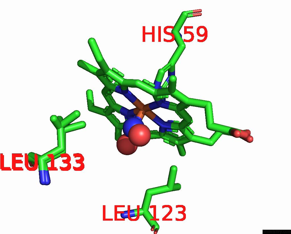
Mono view
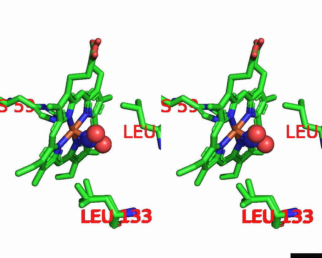
Stereo pair view
Mono view
Stereo pair view
A full contact list of Iron with other atoms in the Fe binding
site number 1 of 1.00 A Crystal Structure of V36A/D129A/L130A Mutant of Nitrophorin 4 From Rhodnius Prolixus Complexed with Nitric Oxide at pH 5.6 within 5.0Å range:
|
Reference:
A.M.Amoia,
W.R.Montfort.
Ligand Protection and Nitric Oxide Interactions with Heme in Nitrophorin 4: Kinetic and Structural Analyses of A Loop Mutant To Be Published.
Page generated: Thu Jul 17 03:12:03 2025
Last articles
Mg in 388DMg in 384D
Mg in 354D
Mg in 383D
Mg in 382D
Mg in 370D
Mg in 360D
Mg in 336D
Mg in 355D
Mg in 308D