Iron »
PDB 2nwf-2oof »
2og6 »
Iron in PDB 2og6: Crystal Structure of Asparagine Oxygenase in Complex with Fe(II)
Protein crystallography data
The structure of Crystal Structure of Asparagine Oxygenase in Complex with Fe(II), PDB code: 2og6
was solved by
L.-O.Essen,
M.Strieker,
with X-Ray Crystallography technique. A brief refinement statistics is given in the table below:
| Resolution Low / High (Å) | 20.00 / 1.92 |
| Space group | P 31 2 1 |
| Cell size a, b, c (Å), α, β, γ (°) | 90.733, 90.733, 89.791, 90.00, 90.00, 120.00 |
| R / Rfree (%) | 16.9 / 19.7 |
Other elements in 2og6:
The structure of Crystal Structure of Asparagine Oxygenase in Complex with Fe(II) also contains other interesting chemical elements:
| Chlorine | (Cl) | 2 atoms |
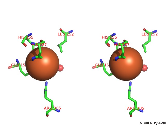
Iron Binding Sites:
The binding sites of Iron atom in the Crystal Structure of Asparagine Oxygenase in Complex with Fe(II)
(pdb code 2og6). This binding sites where shown within
5.0 Angstroms radius around Iron atom.
In total only one binding site of Iron was determined in the Crystal Structure of Asparagine Oxygenase in Complex with Fe(II), PDB code: 2og6:
In total only one binding site of Iron was determined in the Crystal Structure of Asparagine Oxygenase in Complex with Fe(II), PDB code: 2og6:
Iron binding site 1 out of 1 in 2og6
Go back to
Iron binding site 1 out
of 1 in the Crystal Structure of Asparagine Oxygenase in Complex with Fe(II)
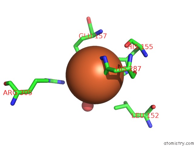
Mono view
Stereo pair view
Mono view
Stereo pair view
A full contact list of Iron with other atoms in the Fe binding
site number 1 of Crystal Structure of Asparagine Oxygenase in Complex with Fe(II) within 5.0Å range:
|
Reference:
M.Strieker,
F.Kopp,
C.Mahlert,
L.-O.Essen,
M.A.Marahiel.
Mechanistic and Structural Basis of Stereospecific Cbeta-Hydroxylation in Calcium-Dependent Antibiotic, A Daptomycin-Type Lipopeptide Acs Chem.Biol. V. 2 187 2007.
ISSN: ISSN 1554-8929
PubMed: 17373765
DOI: 10.1021/CB700012Y
Page generated: Sun Aug 4 00:57:26 2024
ISSN: ISSN 1554-8929
PubMed: 17373765
DOI: 10.1021/CB700012Y
Last articles
Br in 4HSXBr in 4HHQ
Br in 4HHO
Br in 4HFC
Br in 4HH5
Br in 4HC6
Br in 4H2M
Br in 4HBV
Br in 4H9G
Br in 4H5B