Iron »
PDB 2nwf-2oof »
2og7 »
Iron in PDB 2og7: Cystal Structure of Asparagine Oxygenase in Complex with Fe(II), 2S, 3S-3-Hydroxyasparagine and Succinate
Protein crystallography data
The structure of Cystal Structure of Asparagine Oxygenase in Complex with Fe(II), 2S, 3S-3-Hydroxyasparagine and Succinate, PDB code: 2og7
was solved by
L.-O.Essen,
M.Strieker,
with X-Ray Crystallography technique. A brief refinement statistics is given in the table below:
| Resolution Low / High (Å) | 22.91 / 1.66 |
| Space group | P 31 2 1 |
| Cell size a, b, c (Å), α, β, γ (°) | 91.308, 91.308, 90.867, 90.00, 90.00, 120.00 |
| R / Rfree (%) | 16.8 / 18.4 |
Iron Binding Sites:
The binding sites of Iron atom in the Cystal Structure of Asparagine Oxygenase in Complex with Fe(II), 2S, 3S-3-Hydroxyasparagine and Succinate
(pdb code 2og7). This binding sites where shown within
5.0 Angstroms radius around Iron atom.
In total only one binding site of Iron was determined in the Cystal Structure of Asparagine Oxygenase in Complex with Fe(II), 2S, 3S-3-Hydroxyasparagine and Succinate, PDB code: 2og7:
In total only one binding site of Iron was determined in the Cystal Structure of Asparagine Oxygenase in Complex with Fe(II), 2S, 3S-3-Hydroxyasparagine and Succinate, PDB code: 2og7:
Iron binding site 1 out of 1 in 2og7
Go back to
Iron binding site 1 out
of 1 in the Cystal Structure of Asparagine Oxygenase in Complex with Fe(II), 2S, 3S-3-Hydroxyasparagine and Succinate
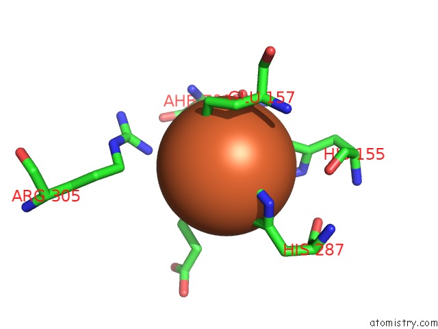
Mono view
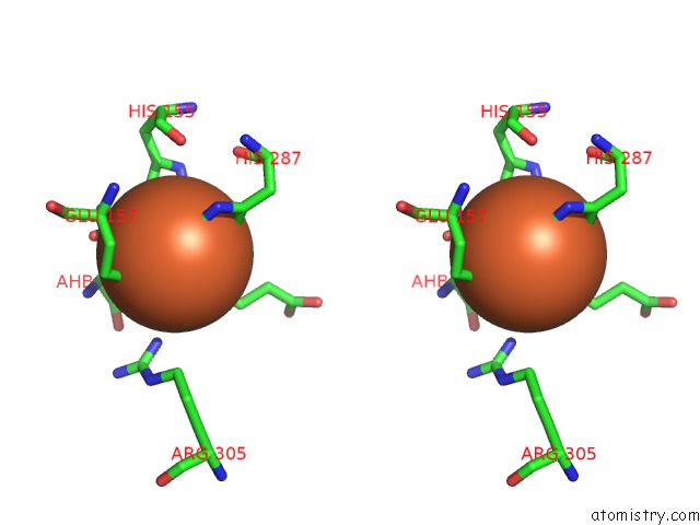
Stereo pair view
Mono view
Stereo pair view
A full contact list of Iron with other atoms in the Fe binding
site number 1 of Cystal Structure of Asparagine Oxygenase in Complex with Fe(II), 2S, 3S-3-Hydroxyasparagine and Succinate within 5.0Å range:
|
Reference:
M.Strieker,
F.Kopp,
C.Mahlert,
L.-O.Essen,
M.A.Marahiel.
Mechanistic and Structural Basis of Stereospecific Cbeta-Hydroxylation in Calcium-Dependent Antibiotic, A Daptomycin-Type Lipopeptide Acs Chem.Biol. V. 2 187 2007.
ISSN: ISSN 1554-8929
PubMed: 17373765
DOI: 10.1021/CB700012Y
Page generated: Thu Jul 17 03:12:11 2025
ISSN: ISSN 1554-8929
PubMed: 17373765
DOI: 10.1021/CB700012Y
Last articles
Mg in 310DMg in 314D
Mg in 313D
Mg in 2ZZN
Mg in 312D
Mg in 306D
Mg in 305D
Mg in 2ZY9
Mg in 301D
Mg in 303D