Iron »
PDB 2orl-2pg7 »
2owt »
Iron in PDB 2owt: Crystal Structure of Bordetella Pertussis Holo Ferric Binding Protein with Bound Synergistic Carbonate Anion
Protein crystallography data
The structure of Crystal Structure of Bordetella Pertussis Holo Ferric Binding Protein with Bound Synergistic Carbonate Anion, PDB code: 2owt
was solved by
S.A.L.Tom-Yew,
M.E.P.Murphy,
with X-Ray Crystallography technique. A brief refinement statistics is given in the table below:
| Resolution Low / High (Å) | 48.00 / 2.40 |
| Space group | P 21 21 21 |
| Cell size a, b, c (Å), α, β, γ (°) | 54.674, 57.278, 87.927, 90.00, 90.00, 90.00 |
| R / Rfree (%) | 17.5 / 24.2 |
Iron Binding Sites:
The binding sites of Iron atom in the Crystal Structure of Bordetella Pertussis Holo Ferric Binding Protein with Bound Synergistic Carbonate Anion
(pdb code 2owt). This binding sites where shown within
5.0 Angstroms radius around Iron atom.
In total only one binding site of Iron was determined in the Crystal Structure of Bordetella Pertussis Holo Ferric Binding Protein with Bound Synergistic Carbonate Anion, PDB code: 2owt:
In total only one binding site of Iron was determined in the Crystal Structure of Bordetella Pertussis Holo Ferric Binding Protein with Bound Synergistic Carbonate Anion, PDB code: 2owt:
Iron binding site 1 out of 1 in 2owt
Go back to
Iron binding site 1 out
of 1 in the Crystal Structure of Bordetella Pertussis Holo Ferric Binding Protein with Bound Synergistic Carbonate Anion
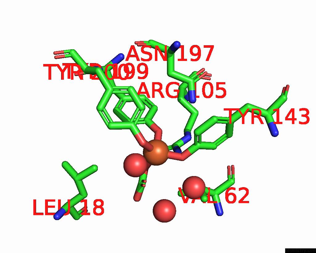
Mono view
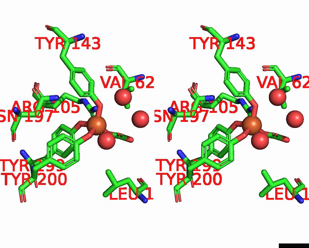
Stereo pair view
Mono view
Stereo pair view
A full contact list of Iron with other atoms in the Fe binding
site number 1 of Crystal Structure of Bordetella Pertussis Holo Ferric Binding Protein with Bound Synergistic Carbonate Anion within 5.0Å range:
|
Reference:
S.A.L.Tom-Yew,
B.H.Shilton,
E.G.Bekker,
E.I.Tocheva,
M.E.P.Murphy.
Anion-Dependent Hinge Motion in Ferric Binding Proteins To Be Published.
Page generated: Thu Jul 17 03:17:09 2025
Last articles
K in 9NESK in 9PHG
K in 9NEI
K in 9NED
K in 9NEC
K in 9NEG
K in 9CWU
K in 9CVB
K in 9CVA
K in 9COM