Iron »
PDB 2orl-2pg7 »
2p1s »
Iron in PDB 2p1s: Crystal Structure of the C-Terminal Lobe of Bovine Lactoferrin Complexed with O-Alpha-D-Glucopyranosyl-(1 3)-Alpha-D- Fructofuranosyl- (2 1)- Alpha-D-Glucopyranoside at 1.93 A Resolution
Protein crystallography data
The structure of Crystal Structure of the C-Terminal Lobe of Bovine Lactoferrin Complexed with O-Alpha-D-Glucopyranosyl-(1 3)-Alpha-D- Fructofuranosyl- (2 1)- Alpha-D-Glucopyranoside at 1.93 A Resolution, PDB code: 2p1s
was solved by
R.Mir,
N.Singh,
M.Sinha,
S.Sharma,
P.Kaur,
T.P.Singh,
with X-Ray Crystallography technique. A brief refinement statistics is given in the table below:
| Resolution Low / High (Å) | 63.25 / 1.93 |
| Space group | P 1 21 1 |
| Cell size a, b, c (Å), α, β, γ (°) | 63.155, 50.425, 65.883, 90.00, 107.70, 90.00 |
| R / Rfree (%) | 17 / 21.7 |
Other elements in 2p1s:
The structure of Crystal Structure of the C-Terminal Lobe of Bovine Lactoferrin Complexed with O-Alpha-D-Glucopyranosyl-(1 3)-Alpha-D- Fructofuranosyl- (2 1)- Alpha-D-Glucopyranoside at 1.93 A Resolution also contains other interesting chemical elements:
| Zinc | (Zn) | 2 atoms |
Iron Binding Sites:
The binding sites of Iron atom in the Crystal Structure of the C-Terminal Lobe of Bovine Lactoferrin Complexed with O-Alpha-D-Glucopyranosyl-(1 3)-Alpha-D- Fructofuranosyl- (2 1)- Alpha-D-Glucopyranoside at 1.93 A Resolution
(pdb code 2p1s). This binding sites where shown within
5.0 Angstroms radius around Iron atom.
In total only one binding site of Iron was determined in the Crystal Structure of the C-Terminal Lobe of Bovine Lactoferrin Complexed with O-Alpha-D-Glucopyranosyl-(1 3)-Alpha-D- Fructofuranosyl- (2 1)- Alpha-D-Glucopyranoside at 1.93 A Resolution, PDB code: 2p1s:
In total only one binding site of Iron was determined in the Crystal Structure of the C-Terminal Lobe of Bovine Lactoferrin Complexed with O-Alpha-D-Glucopyranosyl-(1 3)-Alpha-D- Fructofuranosyl- (2 1)- Alpha-D-Glucopyranoside at 1.93 A Resolution, PDB code: 2p1s:
Iron binding site 1 out of 1 in 2p1s
Go back to
Iron binding site 1 out
of 1 in the Crystal Structure of the C-Terminal Lobe of Bovine Lactoferrin Complexed with O-Alpha-D-Glucopyranosyl-(1 3)-Alpha-D- Fructofuranosyl- (2 1)- Alpha-D-Glucopyranoside at 1.93 A Resolution
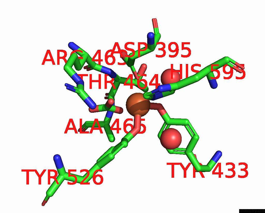
Mono view
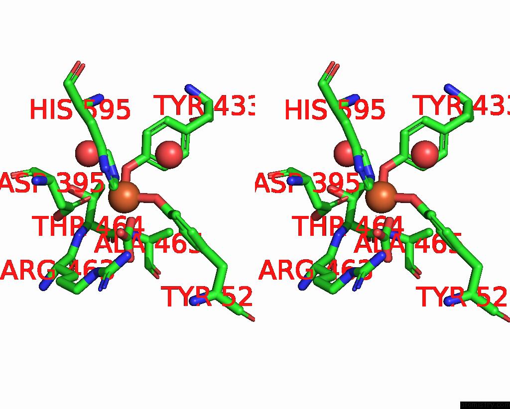
Stereo pair view
Mono view
Stereo pair view
A full contact list of Iron with other atoms in the Fe binding
site number 1 of Crystal Structure of the C-Terminal Lobe of Bovine Lactoferrin Complexed with O-Alpha-D-Glucopyranosyl-(1 3)-Alpha-D- Fructofuranosyl- (2 1)- Alpha-D-Glucopyranoside at 1.93 A Resolution within 5.0Å range:
|
Reference:
R.Mir,
N.Singh,
M.Sinha,
S.Sharma,
P.Kaur,
T.P.Singh.
Crystal Structure of the C-Terminal Lobe of Bovine Lactoferrin Complexed with O-Alpha-D-Glucopyranosyl-(1 3)-Alpha-D-Fructofuranosyl-(2 1)-Alpha-D-Glucopyranoside at 1.93 A Resolution To Be Published.
Page generated: Thu Jul 17 03:21:01 2025
Last articles
Mn in 2GU6Mn in 2GU5
Mn in 2GU4
Mn in 2FFL
Mn in 2GTX
Mn in 2GNM
Mn in 2GND
Mn in 2GMV
Mn in 2GLF
Mn in 2GLK