Iron »
PDB 2orl-2pg7 »
2pa8 »
Iron in PDB 2pa8: X-Ray Crystal Structure of the D/L Subcomplex of the Sulfolobus Solfataricus Rna Polymerase
Enzymatic activity of X-Ray Crystal Structure of the D/L Subcomplex of the Sulfolobus Solfataricus Rna Polymerase
All present enzymatic activity of X-Ray Crystal Structure of the D/L Subcomplex of the Sulfolobus Solfataricus Rna Polymerase:
2.7.7.6;
2.7.7.6;
Protein crystallography data
The structure of X-Ray Crystal Structure of the D/L Subcomplex of the Sulfolobus Solfataricus Rna Polymerase, PDB code: 2pa8
was solved by
A.Hirata,
K.S.Murakami,
with X-Ray Crystallography technique. A brief refinement statistics is given in the table below:
| Resolution Low / High (Å) | 15.00 / 1.76 |
| Space group | I 21 21 21 |
| Cell size a, b, c (Å), α, β, γ (°) | 69.703, 93.346, 128.277, 90.00, 90.00, 90.00 |
| R / Rfree (%) | 21 / 24.7 |
Iron Binding Sites:
The binding sites of Iron atom in the X-Ray Crystal Structure of the D/L Subcomplex of the Sulfolobus Solfataricus Rna Polymerase
(pdb code 2pa8). This binding sites where shown within
5.0 Angstroms radius around Iron atom.
In total 3 binding sites of Iron where determined in the X-Ray Crystal Structure of the D/L Subcomplex of the Sulfolobus Solfataricus Rna Polymerase, PDB code: 2pa8:
Jump to Iron binding site number: 1; 2; 3;
In total 3 binding sites of Iron where determined in the X-Ray Crystal Structure of the D/L Subcomplex of the Sulfolobus Solfataricus Rna Polymerase, PDB code: 2pa8:
Jump to Iron binding site number: 1; 2; 3;
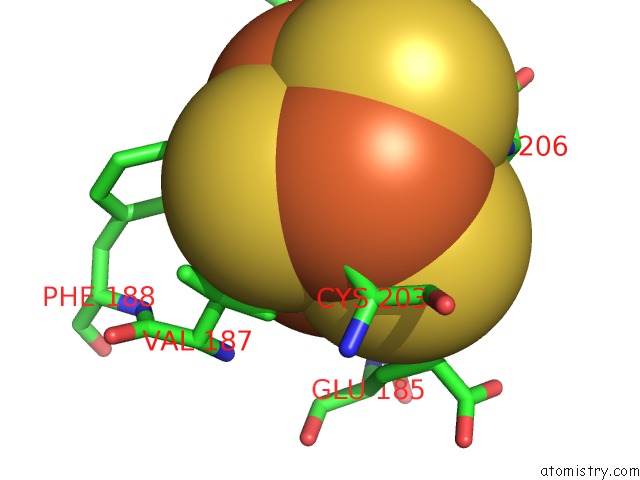
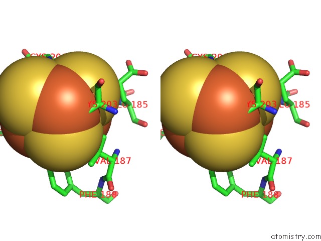
Iron binding site 1 out of 3 in 2pa8
Go back to
Iron binding site 1 out
of 3 in the X-Ray Crystal Structure of the D/L Subcomplex of the Sulfolobus Solfataricus Rna Polymerase
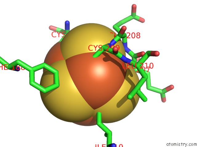
Mono view
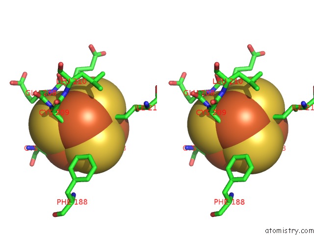
Stereo pair view
Mono view
Stereo pair view
A full contact list of Iron with other atoms in the Fe binding
site number 1 of X-Ray Crystal Structure of the D/L Subcomplex of the Sulfolobus Solfataricus Rna Polymerase within 5.0Å range:
|
Iron binding site 2 out of 3 in 2pa8
Go back to
Iron binding site 2 out
of 3 in the X-Ray Crystal Structure of the D/L Subcomplex of the Sulfolobus Solfataricus Rna Polymerase
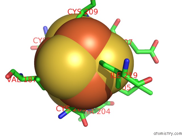
Mono view
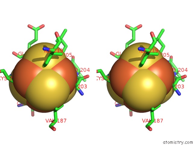
Stereo pair view
Mono view
Stereo pair view
A full contact list of Iron with other atoms in the Fe binding
site number 2 of X-Ray Crystal Structure of the D/L Subcomplex of the Sulfolobus Solfataricus Rna Polymerase within 5.0Å range:
|
Iron binding site 3 out of 3 in 2pa8
Go back to
Iron binding site 3 out
of 3 in the X-Ray Crystal Structure of the D/L Subcomplex of the Sulfolobus Solfataricus Rna Polymerase
Mono view
Stereo pair view
Mono view
Stereo pair view
A full contact list of Iron with other atoms in the Fe binding
site number 3 of X-Ray Crystal Structure of the D/L Subcomplex of the Sulfolobus Solfataricus Rna Polymerase within 5.0Å range:
|
Reference:
A.Hirata,
B.J.Klein,
K.S.Murakami.
The X-Ray Crystal Structure of Rna Polymerase From Archaea. Nature V. 451 851 2008.
ISSN: ISSN 0028-0836
PubMed: 18235446
DOI: 10.1038/NATURE06530
Page generated: Thu Jul 17 03:22:20 2025
ISSN: ISSN 0028-0836
PubMed: 18235446
DOI: 10.1038/NATURE06530
Last articles
K in 9NESK in 9PHG
K in 9NEI
K in 9NED
K in 9NEC
K in 9NEG
K in 9CWU
K in 9CVB
K in 9CVA
K in 9COM