Iron »
PDB 2pgh-2q9u »
2pms »
Iron in PDB 2pms: Crystal Structure of the Complex of Human Lactoferrin N-Lobe and Lactoferrin-Binding Domain of Pneumococcal Surface Protein A
Protein crystallography data
The structure of Crystal Structure of the Complex of Human Lactoferrin N-Lobe and Lactoferrin-Binding Domain of Pneumococcal Surface Protein A, PDB code: 2pms
was solved by
D.Chattopadhyay,
O.Senkovich,
W.J.Cook,
with X-Ray Crystallography technique. A brief refinement statistics is given in the table below:
| Resolution Low / High (Å) | 15.00 / 2.91 |
| Space group | P 32 |
| Cell size a, b, c (Å), α, β, γ (°) | 130.180, 130.180, 80.800, 90.00, 90.00, 120.00 |
| R / Rfree (%) | 20.3 / 24.9 |
Other elements in 2pms:
The structure of Crystal Structure of the Complex of Human Lactoferrin N-Lobe and Lactoferrin-Binding Domain of Pneumococcal Surface Protein A also contains other interesting chemical elements:
| Zinc | (Zn) | 2 atoms |
Iron Binding Sites:
The binding sites of Iron atom in the Crystal Structure of the Complex of Human Lactoferrin N-Lobe and Lactoferrin-Binding Domain of Pneumococcal Surface Protein A
(pdb code 2pms). This binding sites where shown within
5.0 Angstroms radius around Iron atom.
In total 2 binding sites of Iron where determined in the Crystal Structure of the Complex of Human Lactoferrin N-Lobe and Lactoferrin-Binding Domain of Pneumococcal Surface Protein A, PDB code: 2pms:
Jump to Iron binding site number: 1; 2;
In total 2 binding sites of Iron where determined in the Crystal Structure of the Complex of Human Lactoferrin N-Lobe and Lactoferrin-Binding Domain of Pneumococcal Surface Protein A, PDB code: 2pms:
Jump to Iron binding site number: 1; 2;
Iron binding site 1 out of 2 in 2pms
Go back to
Iron binding site 1 out
of 2 in the Crystal Structure of the Complex of Human Lactoferrin N-Lobe and Lactoferrin-Binding Domain of Pneumococcal Surface Protein A
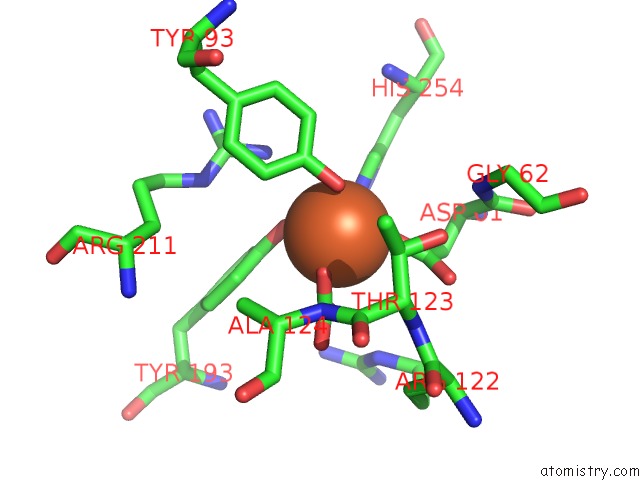
Mono view
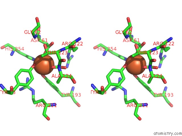
Stereo pair view
Mono view
Stereo pair view
A full contact list of Iron with other atoms in the Fe binding
site number 1 of Crystal Structure of the Complex of Human Lactoferrin N-Lobe and Lactoferrin-Binding Domain of Pneumococcal Surface Protein A within 5.0Å range:
|
Iron binding site 2 out of 2 in 2pms
Go back to
Iron binding site 2 out
of 2 in the Crystal Structure of the Complex of Human Lactoferrin N-Lobe and Lactoferrin-Binding Domain of Pneumococcal Surface Protein A
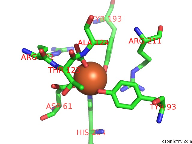
Mono view
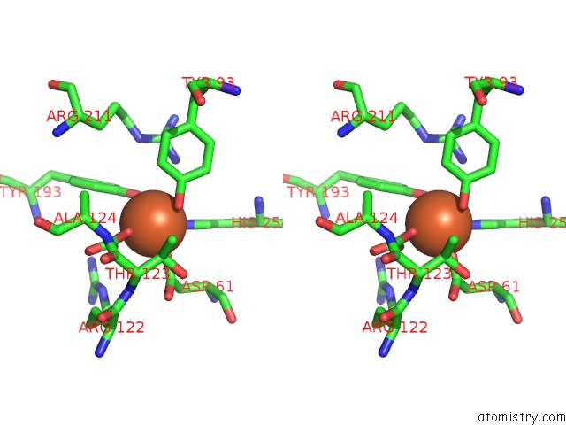
Stereo pair view
Mono view
Stereo pair view
A full contact list of Iron with other atoms in the Fe binding
site number 2 of Crystal Structure of the Complex of Human Lactoferrin N-Lobe and Lactoferrin-Binding Domain of Pneumococcal Surface Protein A within 5.0Å range:
|
Reference:
O.Senkovich,
W.J.Cook,
S.Mirza,
S.K.Hollingshead,
I.I.Protasevich,
D.E.Briles,
D.Chattopadhyay.
Structure of A Complex of Human Lactoferrin N-Lobe with Pneumococcal Surface Protein A Provides Insight Into Microbial Defense Mechanism. J.Mol.Biol. V. 370 701 2007.
ISSN: ISSN 0022-2836
PubMed: 17543335
DOI: 10.1016/J.JMB.2007.04.075
Page generated: Thu Jul 17 03:28:43 2025
ISSN: ISSN 0022-2836
PubMed: 17543335
DOI: 10.1016/J.JMB.2007.04.075
Last articles
Fe in 9AV9Fe in 9AYT
Fe in 8ZWB
Fe in 8ZNO
Fe in 8ZXR
Fe in 8ZXQ
Fe in 8ZJC
Fe in 8ZVH
Fe in 8ZVG
Fe in 8ZNS