Iron »
PDB 2r1m-2rfb »
2r5l »
Iron in PDB 2r5l: Crystal Structure of Lactoperoxidase at 2.4A Resolution
Enzymatic activity of Crystal Structure of Lactoperoxidase at 2.4A Resolution
All present enzymatic activity of Crystal Structure of Lactoperoxidase at 2.4A Resolution:
1.11.1.7;
1.11.1.7;
Protein crystallography data
The structure of Crystal Structure of Lactoperoxidase at 2.4A Resolution, PDB code: 2r5l
was solved by
A.K.Singh,
N.Singh,
S.Sharma,
P.Kaur,
A.Srinivasan,
T.P.Singh,
with X-Ray Crystallography technique. A brief refinement statistics is given in the table below:
| Resolution Low / High (Å) | 19.98 / 2.40 |
| Space group | P 1 21 1 |
| Cell size a, b, c (Å), α, β, γ (°) | 54.190, 80.810, 77.040, 90.00, 102.95, 90.00 |
| R / Rfree (%) | 19.6 / 20.3 |
Other elements in 2r5l:
The structure of Crystal Structure of Lactoperoxidase at 2.4A Resolution also contains other interesting chemical elements:
| Iodine | (I) | 10 atoms |
| Calcium | (Ca) | 1 atom |
Iron Binding Sites:
The binding sites of Iron atom in the Crystal Structure of Lactoperoxidase at 2.4A Resolution
(pdb code 2r5l). This binding sites where shown within
5.0 Angstroms radius around Iron atom.
In total only one binding site of Iron was determined in the Crystal Structure of Lactoperoxidase at 2.4A Resolution, PDB code: 2r5l:
In total only one binding site of Iron was determined in the Crystal Structure of Lactoperoxidase at 2.4A Resolution, PDB code: 2r5l:
Iron binding site 1 out of 1 in 2r5l
Go back to
Iron binding site 1 out
of 1 in the Crystal Structure of Lactoperoxidase at 2.4A Resolution
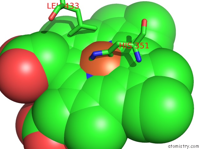
Mono view
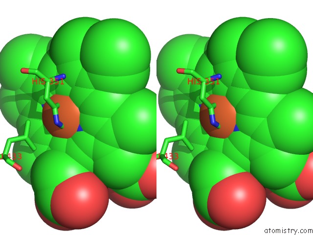
Stereo pair view
Mono view
Stereo pair view
A full contact list of Iron with other atoms in the Fe binding
site number 1 of Crystal Structure of Lactoperoxidase at 2.4A Resolution within 5.0Å range:
|
Reference:
A.K.Singh,
N.Singh,
S.Sharma,
S.B.Singh,
P.Kaur,
A.Bhushan,
A.Srinivasan,
T.P.Singh.
Crystal Structure of Lactoperoxidase at 2.4 A Resolution. J.Mol.Biol. V. 376 1060 2007.
ISSN: ISSN 0022-2836
PubMed: 18191143
DOI: 10.1016/J.JMB.2007.12.012
Page generated: Thu Jul 17 03:54:11 2025
ISSN: ISSN 0022-2836
PubMed: 18191143
DOI: 10.1016/J.JMB.2007.12.012
Last articles
Mg in 1IR1Mg in 1IR3
Mg in 1IQC
Mg in 1IBL
Mg in 1IPW
Mg in 1IQ8
Mg in 1IPP
Mg in 1IOW
Mg in 1IBM
Mg in 1IOV