Iron »
PDB 2r1m-2rfb »
2r6s »
Iron in PDB 2r6s: Crystal Structure of Gab Protein
Protein crystallography data
The structure of Crystal Structure of Gab Protein, PDB code: 2r6s
was solved by
B.Lohkamp,
D.Dobritzsch,
with X-Ray Crystallography technique. A brief refinement statistics is given in the table below:
| Resolution Low / High (Å) | 45.36 / 2.10 |
| Space group | I 4 2 2 |
| Cell size a, b, c (Å), α, β, γ (°) | 120.900, 120.900, 137.170, 90.00, 90.00, 90.00 |
| R / Rfree (%) | 16.2 / 19.9 |
Iron Binding Sites:
The binding sites of Iron atom in the Crystal Structure of Gab Protein
(pdb code 2r6s). This binding sites where shown within
5.0 Angstroms radius around Iron atom.
In total only one binding site of Iron was determined in the Crystal Structure of Gab Protein, PDB code: 2r6s:
In total only one binding site of Iron was determined in the Crystal Structure of Gab Protein, PDB code: 2r6s:
Iron binding site 1 out of 1 in 2r6s
Go back to
Iron binding site 1 out
of 1 in the Crystal Structure of Gab Protein
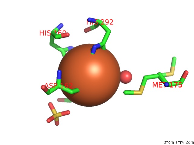
Mono view
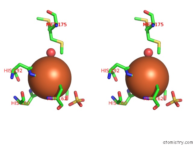
Stereo pair view
Mono view
Stereo pair view
A full contact list of Iron with other atoms in the Fe binding
site number 1 of Crystal Structure of Gab Protein within 5.0Å range:
|
Reference:
B.Lohkamp,
D.Dobritzsch.
A Mixture of Fortunes: the Curious Determination of the Structure of Escherichia Coli BL21 Gab Protein. Acta Crystallogr.,Sect.D V. 64 407 2008.
ISSN: ISSN 0907-4449
PubMed: 18391407
DOI: 10.1107/S0907444908001091
Page generated: Thu Jul 17 03:54:24 2025
ISSN: ISSN 0907-4449
PubMed: 18391407
DOI: 10.1107/S0907444908001091
Last articles
Fe in 2YXOFe in 2YRS
Fe in 2YXC
Fe in 2YNM
Fe in 2YVJ
Fe in 2YP1
Fe in 2YU2
Fe in 2YU1
Fe in 2YQB
Fe in 2YOO