Iron »
PDB 2r1m-2rfb »
2r7a »
Iron in PDB 2r7a: Crystal Structure of A Periplasmic Heme Binding Protein From Shigella Dysenteriae
Protein crystallography data
The structure of Crystal Structure of A Periplasmic Heme Binding Protein From Shigella Dysenteriae, PDB code: 2r7a
was solved by
W.W.Ho,
H.Li,
T.L.Poulos,
with X-Ray Crystallography technique. A brief refinement statistics is given in the table below:
| Resolution Low / High (Å) | 43.86 / 2.05 |
| Space group | P 1 |
| Cell size a, b, c (Å), α, β, γ (°) | 54.700, 73.308, 73.316, 70.25, 79.02, 90.22 |
| R / Rfree (%) | 21 / 25.5 |
Iron Binding Sites:
The binding sites of Iron atom in the Crystal Structure of A Periplasmic Heme Binding Protein From Shigella Dysenteriae
(pdb code 2r7a). This binding sites where shown within
5.0 Angstroms radius around Iron atom.
In total only one binding site of Iron was determined in the Crystal Structure of A Periplasmic Heme Binding Protein From Shigella Dysenteriae, PDB code: 2r7a:
In total only one binding site of Iron was determined in the Crystal Structure of A Periplasmic Heme Binding Protein From Shigella Dysenteriae, PDB code: 2r7a:
Iron binding site 1 out of 1 in 2r7a
Go back to
Iron binding site 1 out
of 1 in the Crystal Structure of A Periplasmic Heme Binding Protein From Shigella Dysenteriae
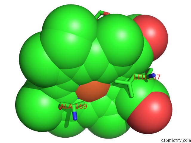
Mono view
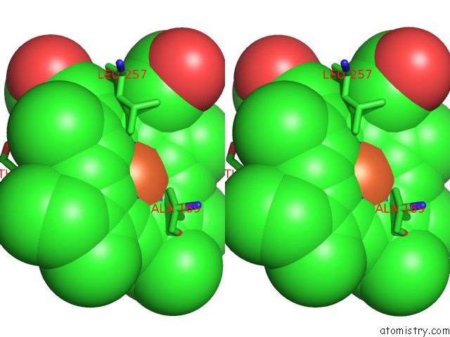
Stereo pair view
Mono view
Stereo pair view
A full contact list of Iron with other atoms in the Fe binding
site number 1 of Crystal Structure of A Periplasmic Heme Binding Protein From Shigella Dysenteriae within 5.0Å range:
|
Reference:
W.W.Ho,
H.Li,
S.Eakanunkul,
Y.Tong,
A.Wilks,
M.Guo,
T.L.Poulos.
Holo- and Apo-Bound Structures of Bacterial Periplasmic Heme-Binding Proteins. J.Biol.Chem. V. 282 35796 2007.
ISSN: ISSN 0021-9258
PubMed: 17925389
DOI: 10.1074/JBC.M706761200
Page generated: Thu Jul 17 03:54:44 2025
ISSN: ISSN 0021-9258
PubMed: 17925389
DOI: 10.1074/JBC.M706761200
Last articles
Fe in 2YXOFe in 2YRS
Fe in 2YXC
Fe in 2YNM
Fe in 2YVJ
Fe in 2YP1
Fe in 2YU2
Fe in 2YU1
Fe in 2YQB
Fe in 2YOO