Iron »
PDB 2r1m-2rfb »
2r9j »
Iron in PDB 2r9j: Ligand Recognition in C-Lobe: the Crystal Structure of the Complex of Lactoferrin C-Lobe with Nicotinamide at 2.5 A Resolution
Protein crystallography data
The structure of Ligand Recognition in C-Lobe: the Crystal Structure of the Complex of Lactoferrin C-Lobe with Nicotinamide at 2.5 A Resolution, PDB code: 2r9j
was solved by
R.Mir,
G.Vikram,
N.Singh,
S.Kumar,
M.Sinha,
S.Sharma,
P.Kaur,
T.P.Singh,
with X-Ray Crystallography technique. A brief refinement statistics is given in the table below:
| Resolution Low / High (Å) | 20.00 / 2.55 |
| Space group | P 1 21 1 |
| Cell size a, b, c (Å), α, β, γ (°) | 63.330, 50.340, 65.910, 90.00, 107.65, 90.00 |
| R / Rfree (%) | 22.5 / 24.9 |
Other elements in 2r9j:
The structure of Ligand Recognition in C-Lobe: the Crystal Structure of the Complex of Lactoferrin C-Lobe with Nicotinamide at 2.5 A Resolution also contains other interesting chemical elements:
| Zinc | (Zn) | 2 atoms |
Iron Binding Sites:
The binding sites of Iron atom in the Ligand Recognition in C-Lobe: the Crystal Structure of the Complex of Lactoferrin C-Lobe with Nicotinamide at 2.5 A Resolution
(pdb code 2r9j). This binding sites where shown within
5.0 Angstroms radius around Iron atom.
In total only one binding site of Iron was determined in the Ligand Recognition in C-Lobe: the Crystal Structure of the Complex of Lactoferrin C-Lobe with Nicotinamide at 2.5 A Resolution, PDB code: 2r9j:
In total only one binding site of Iron was determined in the Ligand Recognition in C-Lobe: the Crystal Structure of the Complex of Lactoferrin C-Lobe with Nicotinamide at 2.5 A Resolution, PDB code: 2r9j:
Iron binding site 1 out of 1 in 2r9j
Go back to
Iron binding site 1 out
of 1 in the Ligand Recognition in C-Lobe: the Crystal Structure of the Complex of Lactoferrin C-Lobe with Nicotinamide at 2.5 A Resolution

Mono view
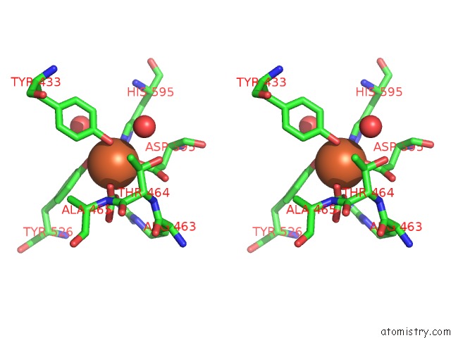
Stereo pair view
Mono view
Stereo pair view
A full contact list of Iron with other atoms in the Fe binding
site number 1 of Ligand Recognition in C-Lobe: the Crystal Structure of the Complex of Lactoferrin C-Lobe with Nicotinamide at 2.5 A Resolution within 5.0Å range:
|
Reference:
R.Mir,
G.Vikram,
N.Singh,
S.Kumar,
M.Sinha,
S.Sharma,
P.Kaur,
T.P.Singh.
Ligand Recognition in C-Lobe: the Crystal Structure of the Complex of Lactoferrin C-Lobe with Nicotinamide at 2.5 A Resolution To Be Published.
Page generated: Thu Jul 17 03:54:57 2025
Last articles
Mn in 3VNJMn in 3VNI
Mn in 3VET
Mn in 3VES
Mn in 3V9D
Mn in 3V91
Mn in 3V0Q
Mn in 3V1R
Mn in 3V8Z
Mn in 3V0P