Iron »
PDB 2rfc-2v1i »
2uwv »
Iron in PDB 2uwv: X-Ray High Resolution Structure of the Photosynthetic Reaction Center From Rb. Sphaeroides at pH 6.5 in the Charge-Separated State, 3RD Dataset
Protein crystallography data
The structure of X-Ray High Resolution Structure of the Photosynthetic Reaction Center From Rb. Sphaeroides at pH 6.5 in the Charge-Separated State, 3RD Dataset, PDB code: 2uwv
was solved by
J.Koepke,
R.Diehm,
G.Fritzsch,
with X-Ray Crystallography technique. A brief refinement statistics is given in the table below:
| Resolution Low / High (Å) | 119.52 / 2.13 |
| Space group | P 31 2 1 |
| Cell size a, b, c (Å), α, β, γ (°) | 139.662, 139.662, 184.436, 90.00, 90.00, 120.00 |
| R / Rfree (%) | 22.5 / 24.7 |
Other elements in 2uwv:
The structure of X-Ray High Resolution Structure of the Photosynthetic Reaction Center From Rb. Sphaeroides at pH 6.5 in the Charge-Separated State, 3RD Dataset also contains other interesting chemical elements:
| Magnesium | (Mg) | 4 atoms |
Iron Binding Sites:
The binding sites of Iron atom in the X-Ray High Resolution Structure of the Photosynthetic Reaction Center From Rb. Sphaeroides at pH 6.5 in the Charge-Separated State, 3RD Dataset
(pdb code 2uwv). This binding sites where shown within
5.0 Angstroms radius around Iron atom.
In total only one binding site of Iron was determined in the X-Ray High Resolution Structure of the Photosynthetic Reaction Center From Rb. Sphaeroides at pH 6.5 in the Charge-Separated State, 3RD Dataset, PDB code: 2uwv:
In total only one binding site of Iron was determined in the X-Ray High Resolution Structure of the Photosynthetic Reaction Center From Rb. Sphaeroides at pH 6.5 in the Charge-Separated State, 3RD Dataset, PDB code: 2uwv:
Iron binding site 1 out of 1 in 2uwv
Go back to
Iron binding site 1 out
of 1 in the X-Ray High Resolution Structure of the Photosynthetic Reaction Center From Rb. Sphaeroides at pH 6.5 in the Charge-Separated State, 3RD Dataset
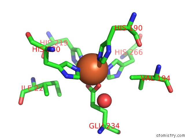
Mono view
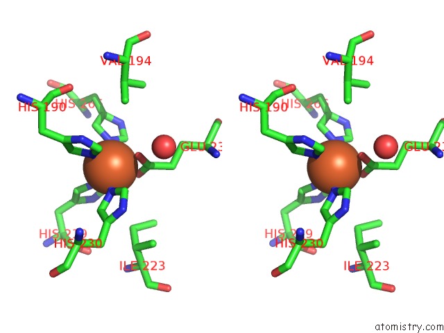
Stereo pair view
Mono view
Stereo pair view
A full contact list of Iron with other atoms in the Fe binding
site number 1 of X-Ray High Resolution Structure of the Photosynthetic Reaction Center From Rb. Sphaeroides at pH 6.5 in the Charge-Separated State, 3RD Dataset within 5.0Å range:
|
Reference:
J.Koepke,
E.M.Krammer,
A.R.Klingen,
P.Sebban,
G.M.Ullmann,
G.Fritzsch.
pH Modulates the Quinone Position in the Photosynthetic Reaction Center From Rhodobacter Sphaeroides in the Neutral and Charge Separated States. J.Mol.Biol. V. 371 396 2007.
ISSN: ISSN 0022-2836
PubMed: 17570397
DOI: 10.1016/J.JMB.2007.04.082
Page generated: Thu Jul 17 04:04:15 2025
ISSN: ISSN 0022-2836
PubMed: 17570397
DOI: 10.1016/J.JMB.2007.04.082
Last articles
Mg in 4DUYMg in 4DR7
Mg in 4DR6
Mg in 4DR5
Mg in 4DUX
Mg in 4DUW
Mg in 4DUV
Mg in 4DUO
Mg in 4DUG
Mg in 4DTY