Iron »
PDB 2w3h-2wl3 »
2w6y »
Iron in PDB 2w6y: Crystal Structure of Sperm Whale Myoglobin Mutant Yqr in Complex with Xenon
Protein crystallography data
The structure of Crystal Structure of Sperm Whale Myoglobin Mutant Yqr in Complex with Xenon, PDB code: 2w6y
was solved by
A.E.Miele,
F.Draghi,
F.Renzi,
G.Sciara,
K.A.Johnson,
B.Vallone,
M.Brunori,
C.Savino,
with X-Ray Crystallography technique. A brief refinement statistics is given in the table below:
| Resolution Low / High (Å) | 26.00 / 1.60 |
| Space group | P 6 |
| Cell size a, b, c (Å), α, β, γ (°) | 90.626, 90.626, 45.505, 90.00, 90.00, 120.00 |
| R / Rfree (%) | 16.5 / 19.6 |
Other elements in 2w6y:
The structure of Crystal Structure of Sperm Whale Myoglobin Mutant Yqr in Complex with Xenon also contains other interesting chemical elements:
| Xenon | (Xe) | 4 atoms |
Iron Binding Sites:
The binding sites of Iron atom in the Crystal Structure of Sperm Whale Myoglobin Mutant Yqr in Complex with Xenon
(pdb code 2w6y). This binding sites where shown within
5.0 Angstroms radius around Iron atom.
In total only one binding site of Iron was determined in the Crystal Structure of Sperm Whale Myoglobin Mutant Yqr in Complex with Xenon, PDB code: 2w6y:
In total only one binding site of Iron was determined in the Crystal Structure of Sperm Whale Myoglobin Mutant Yqr in Complex with Xenon, PDB code: 2w6y:
Iron binding site 1 out of 1 in 2w6y
Go back to
Iron binding site 1 out
of 1 in the Crystal Structure of Sperm Whale Myoglobin Mutant Yqr in Complex with Xenon
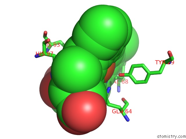
Mono view
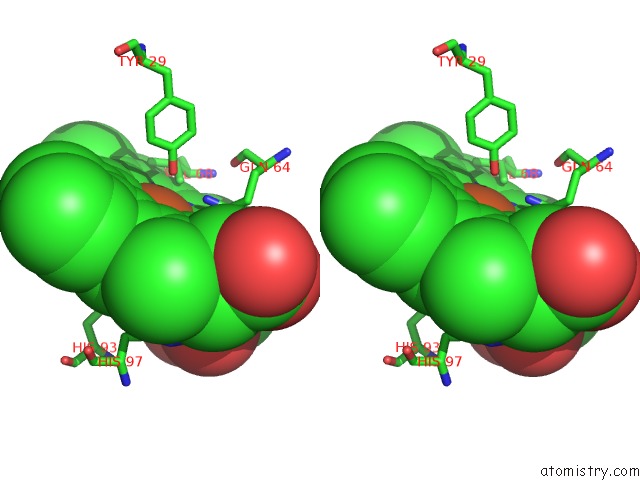
Stereo pair view
Mono view
Stereo pair view
A full contact list of Iron with other atoms in the Fe binding
site number 1 of Crystal Structure of Sperm Whale Myoglobin Mutant Yqr in Complex with Xenon within 5.0Å range:
|
Reference:
A.E.Miele,
F.Draghi,
K.A.Johnson,
F.Renzi,
G.Sciara,
M.Brunori,
B.Vallone,
C.Savino.
When the Same Fold Does Not Mean the Same Function: the Case of Xenon Cavities in Hemoglobin and Myoglobin To Be Published.
Page generated: Thu Jul 17 04:56:57 2025
Last articles
Zn in 1XAGZn in 1X8H
Zn in 1XA6
Zn in 1X8I
Zn in 1X6M
Zn in 1X8G
Zn in 1X6H
Zn in 1X81
Zn in 1X6E
Zn in 1X6F