Iron »
PDB 2w3h-2wl3 »
2wci »
Iron in PDB 2wci: Structure of E. Coli Monothiol Glutaredoxin GRX4 Homodimer
Protein crystallography data
The structure of Structure of E. Coli Monothiol Glutaredoxin GRX4 Homodimer, PDB code: 2wci
was solved by
T.Iwema,
A.Picchiocci,
D.A.K.Traore,
J.-L.Ferrer,
F.Chauvat,
L.Jacquamet,
with X-Ray Crystallography technique. A brief refinement statistics is given in the table below:
| Resolution Low / High (Å) | 47.14 / 1.90 |
| Space group | P 43 21 2 |
| Cell size a, b, c (Å), α, β, γ (°) | 94.300, 94.300, 62.660, 90.00, 90.00, 90.00 |
| R / Rfree (%) | 18.3 / 23.4 |
Other elements in 2wci:
The structure of Structure of E. Coli Monothiol Glutaredoxin GRX4 Homodimer also contains other interesting chemical elements:
| Sodium | (Na) | 1 atom |
Iron Binding Sites:
The binding sites of Iron atom in the Structure of E. Coli Monothiol Glutaredoxin GRX4 Homodimer
(pdb code 2wci). This binding sites where shown within
5.0 Angstroms radius around Iron atom.
In total 2 binding sites of Iron where determined in the Structure of E. Coli Monothiol Glutaredoxin GRX4 Homodimer, PDB code: 2wci:
Jump to Iron binding site number: 1; 2;
In total 2 binding sites of Iron where determined in the Structure of E. Coli Monothiol Glutaredoxin GRX4 Homodimer, PDB code: 2wci:
Jump to Iron binding site number: 1; 2;
Iron binding site 1 out of 2 in 2wci
Go back to
Iron binding site 1 out
of 2 in the Structure of E. Coli Monothiol Glutaredoxin GRX4 Homodimer
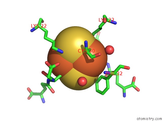
Mono view
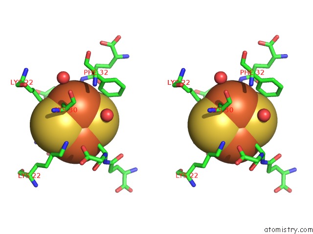
Stereo pair view
Mono view
Stereo pair view
|
|
A full contact list of Iron with other atoms in the Fe binding
site number 1 of Structure of E. Coli Monothiol Glutaredoxin GRX4 Homodimer within 5.0Å range:
|
Iron binding site 2 out of 2 in 2wci
Go back to
Iron binding site 2 out
of 2 in the Structure of E. Coli Monothiol Glutaredoxin GRX4 Homodimer
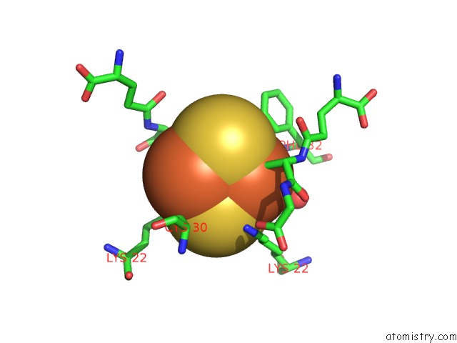
Mono view
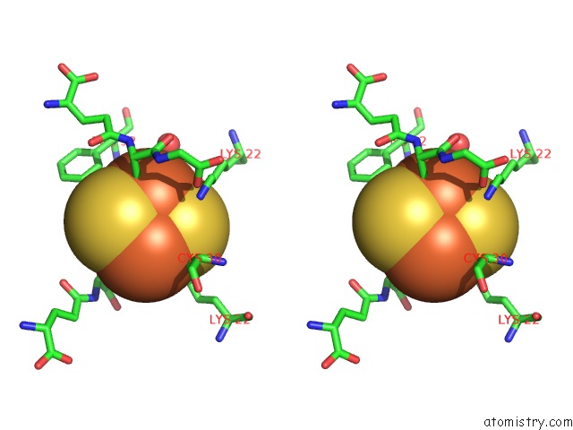
Stereo pair view
Mono view
Stereo pair view
|
|
A full contact list of Iron with other atoms in the Fe binding
site number 2 of Structure of E. Coli Monothiol Glutaredoxin GRX4 Homodimer within 5.0Å range:
|
Reference:
T.Iwema,
A.Picciocchi,
D.A.K.Traore,
J.-L.Ferrer,
F.Chauvat,
L.Jacquamet.
Structural Basis For Delivery of the Intact [FE2S2] Cluster By Monothiol Glutaredoxin. Biochemistry V. 48 6041 2009.
ISSN: ISSN 0006-2960
PubMed: 19505088
DOI: 10.1021/BI900440M
Page generated: Thu Jul 17 04:58:51 2025
ISSN: ISSN 0006-2960
PubMed: 19505088
DOI: 10.1021/BI900440M
Last articles
Zn in 9QM9Zn in 9S44
Zn in 9OFE
Zn in 9OFC
Zn in 9OFD
Zn in 9OF1
Zn in 9OFB
Zn in 9N0J
Zn in 9M5X
Zn in 9LGI