Iron »
PDB 2xf2-2xuz »
2xr8 »
Iron in PDB 2xr8: Crystal Structure of Biphenyl Dioxygenase From Burkholderia Xenovorans LB400
Enzymatic activity of Crystal Structure of Biphenyl Dioxygenase From Burkholderia Xenovorans LB400
All present enzymatic activity of Crystal Structure of Biphenyl Dioxygenase From Burkholderia Xenovorans LB400:
1.14.12.18;
1.14.12.18;
Protein crystallography data
The structure of Crystal Structure of Biphenyl Dioxygenase From Burkholderia Xenovorans LB400, PDB code: 2xr8
was solved by
P.Kumar,
J.T.Bolin,
with X-Ray Crystallography technique. A brief refinement statistics is given in the table below:
| Resolution Low / High (Å) | 129.10 / 2.49 |
| Space group | P 1 |
| Cell size a, b, c (Å), α, β, γ (°) | 132.585, 132.350, 132.984, 102.60, 102.68, 104.61 |
| R / Rfree (%) | 21.633 / 26.714 |
Iron Binding Sites:
Pages:
>>> Page 1 <<< Page 2, Binding sites: 11 - 20; Page 3, Binding sites: 21 - 30; Page 4, Binding sites: 31 - 36;Binding sites:
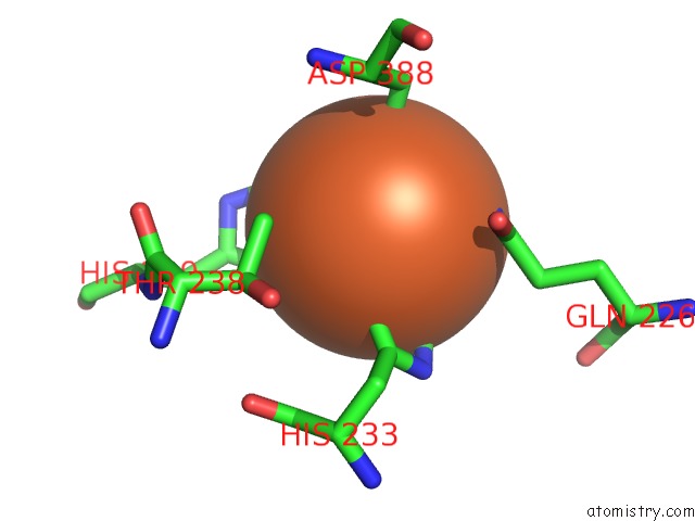
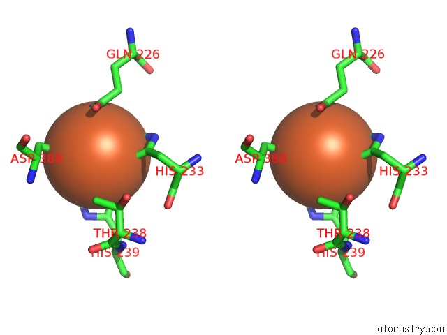
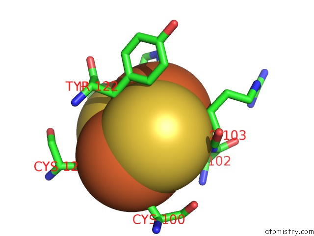
The binding sites of Iron atom in the Crystal Structure of Biphenyl Dioxygenase From Burkholderia Xenovorans LB400 (pdb code 2xr8). This binding sites where shown within 5.0 Angstroms radius around Iron atom.In total 36 binding sites of Iron where determined in the Crystal Structure of Biphenyl Dioxygenase From Burkholderia Xenovorans LB400, PDB code: 2xr8:
Jump to Iron binding site number: 1; 2; 3; 4; 5; 6; 7; 8; 9; 10;
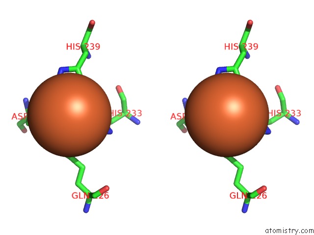
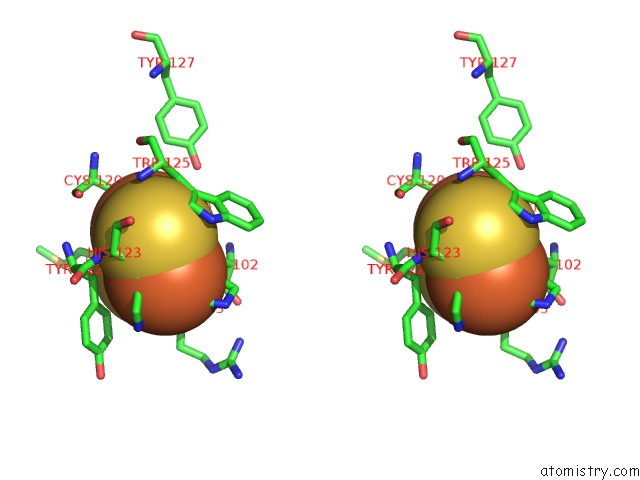
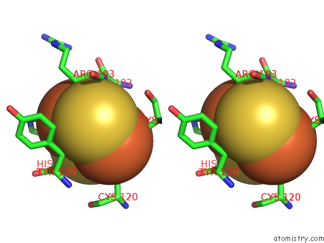
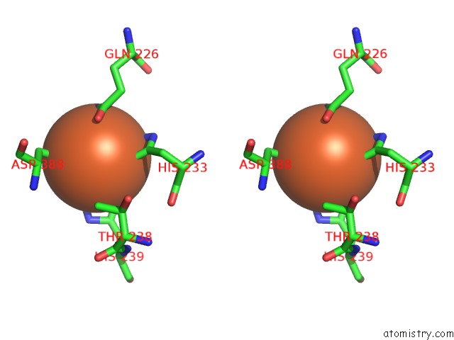
Iron binding site 1 out of 36 in 2xr8
Go back to
Iron binding site 1 out
of 36 in the Crystal Structure of Biphenyl Dioxygenase From Burkholderia Xenovorans LB400
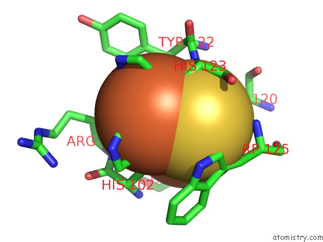
Mono view
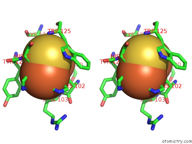
Stereo pair view
Mono view
Stereo pair view
A full contact list of Iron with other atoms in the Fe binding
site number 1 of Crystal Structure of Biphenyl Dioxygenase From Burkholderia Xenovorans LB400 within 5.0Å range:
|
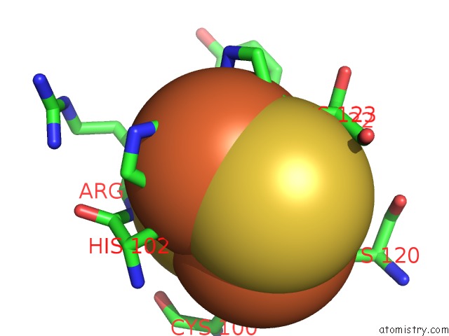
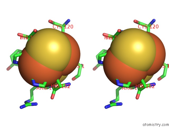
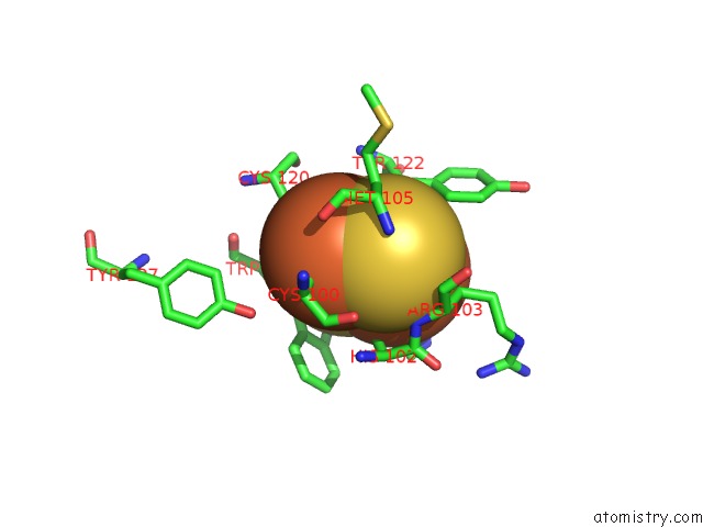
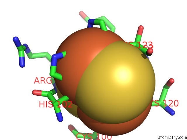
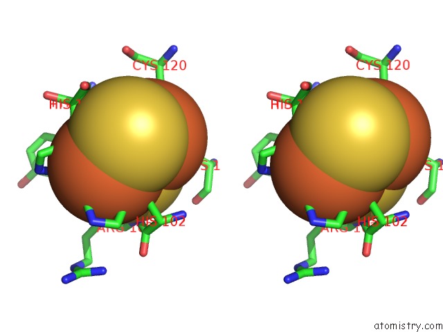
Iron binding site 2 out of 36 in 2xr8
Go back to
Iron binding site 2 out
of 36 in the Crystal Structure of Biphenyl Dioxygenase From Burkholderia Xenovorans LB400
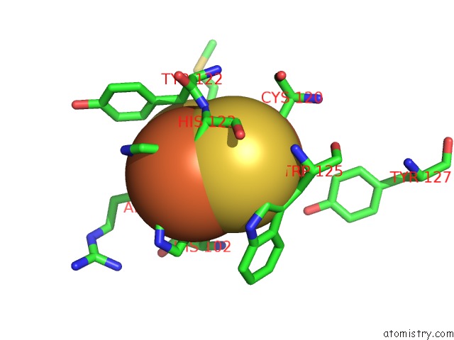
Mono view

Stereo pair view
Mono view
Stereo pair view
A full contact list of Iron with other atoms in the Fe binding
site number 2 of Crystal Structure of Biphenyl Dioxygenase From Burkholderia Xenovorans LB400 within 5.0Å range:
|
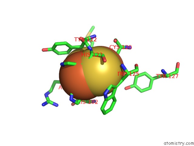
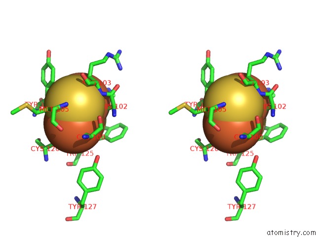
Iron binding site 3 out of 36 in 2xr8
Go back to
Iron binding site 3 out
of 36 in the Crystal Structure of Biphenyl Dioxygenase From Burkholderia Xenovorans LB400
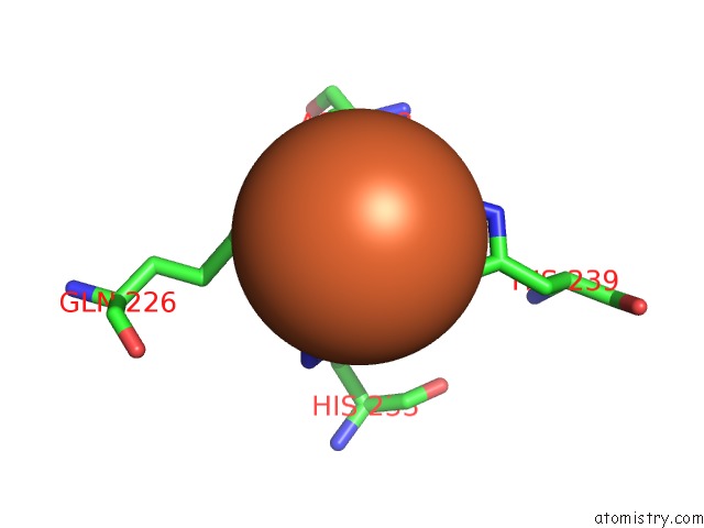
Mono view
Stereo pair view
Mono view
Stereo pair view
A full contact list of Iron with other atoms in the Fe binding
site number 3 of Crystal Structure of Biphenyl Dioxygenase From Burkholderia Xenovorans LB400 within 5.0Å range:
|
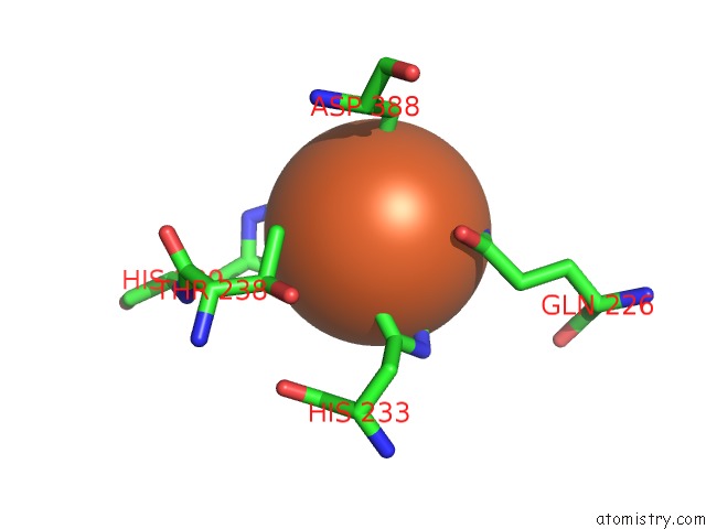
Iron binding site 4 out of 36 in 2xr8
Go back to
Iron binding site 4 out
of 36 in the Crystal Structure of Biphenyl Dioxygenase From Burkholderia Xenovorans LB400
Mono view
Stereo pair view
Mono view
Stereo pair view
A full contact list of Iron with other atoms in the Fe binding
site number 4 of Crystal Structure of Biphenyl Dioxygenase From Burkholderia Xenovorans LB400 within 5.0Å range:
|
Iron binding site 5 out of 36 in 2xr8
Go back to
Iron binding site 5 out
of 36 in the Crystal Structure of Biphenyl Dioxygenase From Burkholderia Xenovorans LB400
Mono view
Stereo pair view
Mono view
Stereo pair view
A full contact list of Iron with other atoms in the Fe binding
site number 5 of Crystal Structure of Biphenyl Dioxygenase From Burkholderia Xenovorans LB400 within 5.0Å range:
|
Iron binding site 6 out of 36 in 2xr8
Go back to
Iron binding site 6 out
of 36 in the Crystal Structure of Biphenyl Dioxygenase From Burkholderia Xenovorans LB400
Mono view
Stereo pair view
Mono view
Stereo pair view
A full contact list of Iron with other atoms in the Fe binding
site number 6 of Crystal Structure of Biphenyl Dioxygenase From Burkholderia Xenovorans LB400 within 5.0Å range:
|
Iron binding site 7 out of 36 in 2xr8
Go back to
Iron binding site 7 out
of 36 in the Crystal Structure of Biphenyl Dioxygenase From Burkholderia Xenovorans LB400
Mono view
Stereo pair view
Mono view
Stereo pair view
A full contact list of Iron with other atoms in the Fe binding
site number 7 of Crystal Structure of Biphenyl Dioxygenase From Burkholderia Xenovorans LB400 within 5.0Å range:
|
Iron binding site 8 out of 36 in 2xr8
Go back to
Iron binding site 8 out
of 36 in the Crystal Structure of Biphenyl Dioxygenase From Burkholderia Xenovorans LB400
Mono view
Stereo pair view
Mono view
Stereo pair view
A full contact list of Iron with other atoms in the Fe binding
site number 8 of Crystal Structure of Biphenyl Dioxygenase From Burkholderia Xenovorans LB400 within 5.0Å range:
|
Iron binding site 9 out of 36 in 2xr8
Go back to
Iron binding site 9 out
of 36 in the Crystal Structure of Biphenyl Dioxygenase From Burkholderia Xenovorans LB400
Mono view
Stereo pair view
Mono view
Stereo pair view
A full contact list of Iron with other atoms in the Fe binding
site number 9 of Crystal Structure of Biphenyl Dioxygenase From Burkholderia Xenovorans LB400 within 5.0Å range:
|
Iron binding site 10 out of 36 in 2xr8
Go back to
Iron binding site 10 out
of 36 in the Crystal Structure of Biphenyl Dioxygenase From Burkholderia Xenovorans LB400
Mono view
Stereo pair view
Mono view
Stereo pair view
A full contact list of Iron with other atoms in the Fe binding
site number 10 of Crystal Structure of Biphenyl Dioxygenase From Burkholderia Xenovorans LB400 within 5.0Å range:
|
Reference:
P.Kumar,
M.Mohammadi,
J.F.Viger,
D.Barriault,
L.Gomez-Gil,
L.D.Eltis,
J.T.Bolin,
M.Sylvestre.
Structural Insight Into the Expanded Pcb-Degrading Abilities of A Biphenyl Dioxygenase Obtained By Directed Evolution. J.Mol.Biol. V. 405 531 2011.
ISSN: ISSN 0022-2836
PubMed: 21073881
DOI: 10.1016/J.JMB.2010.11.009
Page generated: Thu Jul 17 05:35:00 2025
ISSN: ISSN 0022-2836
PubMed: 21073881
DOI: 10.1016/J.JMB.2010.11.009
Last articles
Na in 6TS4Na in 6TRR
Na in 6TQD
Na in 6TQQ
Na in 6TQP
Na in 6TQB
Na in 6TP2
Na in 6TP0
Na in 6TP1
Na in 6TQ4