Iron »
PDB 2xv1-2yde »
2y5a »
Iron in PDB 2y5a: Cytochrome C Peroxidase (Ccp) W191G Bound to 3-Aminopyridine
Enzymatic activity of Cytochrome C Peroxidase (Ccp) W191G Bound to 3-Aminopyridine
All present enzymatic activity of Cytochrome C Peroxidase (Ccp) W191G Bound to 3-Aminopyridine:
1.11.1.5;
1.11.1.5;
Protein crystallography data
The structure of Cytochrome C Peroxidase (Ccp) W191G Bound to 3-Aminopyridine, PDB code: 2y5a
was solved by
D.Cappel,
R.Wahlstrom,
R.Brenk,
C.A.Sotriffer,
with X-Ray Crystallography technique. A brief refinement statistics is given in the table below:
| Resolution Low / High (Å) | 25.18 / 1.25 |
| Space group | P 21 21 21 |
| Cell size a, b, c (Å), α, β, γ (°) | 50.853, 75.362, 106.884, 90.00, 90.00, 90.00 |
| R / Rfree (%) | 14.952 / 16.388 |
Iron Binding Sites:
The binding sites of Iron atom in the Cytochrome C Peroxidase (Ccp) W191G Bound to 3-Aminopyridine
(pdb code 2y5a). This binding sites where shown within
5.0 Angstroms radius around Iron atom.
In total only one binding site of Iron was determined in the Cytochrome C Peroxidase (Ccp) W191G Bound to 3-Aminopyridine, PDB code: 2y5a:
In total only one binding site of Iron was determined in the Cytochrome C Peroxidase (Ccp) W191G Bound to 3-Aminopyridine, PDB code: 2y5a:
Iron binding site 1 out of 1 in 2y5a
Go back to
Iron binding site 1 out
of 1 in the Cytochrome C Peroxidase (Ccp) W191G Bound to 3-Aminopyridine
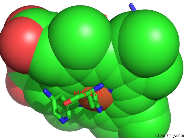
Mono view
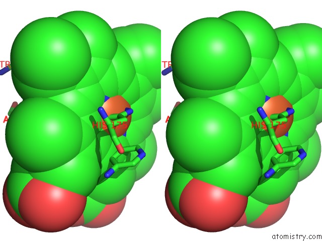
Stereo pair view
Mono view
Stereo pair view
A full contact list of Iron with other atoms in the Fe binding
site number 1 of Cytochrome C Peroxidase (Ccp) W191G Bound to 3-Aminopyridine within 5.0Å range:
|
Reference:
D.Cappel,
R.Wahlstrom,
R.Brenk,
C.A.Sotriffer.
Probing the Dynamic Nature of Water Molecules and Their Influences on Ligand Binding in A Model Binding Site. J.Chem.Inf.Model V. 51 2581 2011.
ISSN: ISSN 1549-9596
PubMed: 21916516
DOI: 10.1021/CI200052J
Page generated: Thu Jul 17 05:52:50 2025
ISSN: ISSN 1549-9596
PubMed: 21916516
DOI: 10.1021/CI200052J
Last articles
Mg in 1VPAMg in 1VPE
Mg in 1VOM
Mg in 1VMA
Mg in 1VMK
Mg in 1VM9
Mg in 1VCR
Mg in 1VLB
Mg in 1VKP
Mg in 1VL8