Iron »
PDB 2z5z-2zpg »
2zfo »
Iron in PDB 2zfo: Structure of the Partially Unliganded Met State of 400 kDa Hemoglobin: Insights Into Ligand-Induced Structural Changes of Giant Hemoglobins
Protein crystallography data
The structure of Structure of the Partially Unliganded Met State of 400 kDa Hemoglobin: Insights Into Ligand-Induced Structural Changes of Giant Hemoglobins, PDB code: 2zfo
was solved by
N.Numoto,
T.Nakagawa,
A.Kita,
Y.Sasayama,
Y.Fukumori,
K.Miki,
with X-Ray Crystallography technique. A brief refinement statistics is given in the table below:
| Resolution Low / High (Å) | 39.22 / 1.95 |
| Space group | H 3 2 |
| Cell size a, b, c (Å), α, β, γ (°) | 110.960, 110.960, 271.580, 90.00, 90.00, 120.00 |
| R / Rfree (%) | 16.9 / 20.2 |
Iron Binding Sites:
The binding sites of Iron atom in the Structure of the Partially Unliganded Met State of 400 kDa Hemoglobin: Insights Into Ligand-Induced Structural Changes of Giant Hemoglobins
(pdb code 2zfo). This binding sites where shown within
5.0 Angstroms radius around Iron atom.
In total 4 binding sites of Iron where determined in the Structure of the Partially Unliganded Met State of 400 kDa Hemoglobin: Insights Into Ligand-Induced Structural Changes of Giant Hemoglobins, PDB code: 2zfo:
Jump to Iron binding site number: 1; 2; 3; 4;
In total 4 binding sites of Iron where determined in the Structure of the Partially Unliganded Met State of 400 kDa Hemoglobin: Insights Into Ligand-Induced Structural Changes of Giant Hemoglobins, PDB code: 2zfo:
Jump to Iron binding site number: 1; 2; 3; 4;
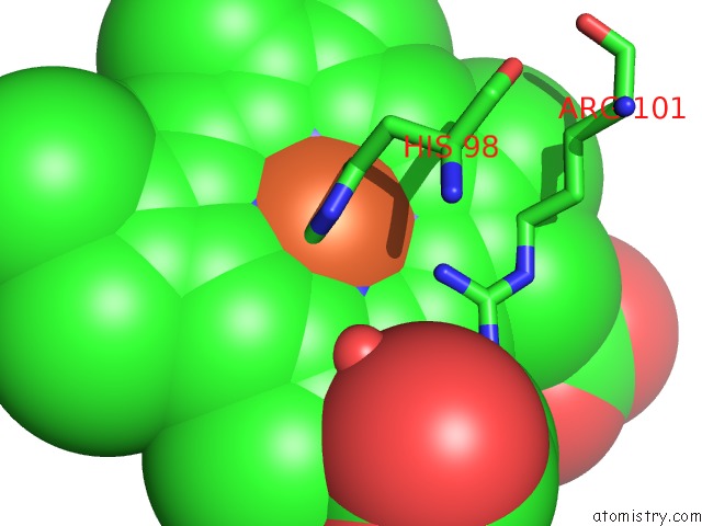
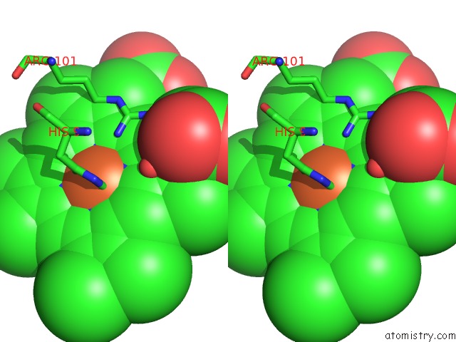
Iron binding site 1 out of 4 in 2zfo
Go back to
Iron binding site 1 out
of 4 in the Structure of the Partially Unliganded Met State of 400 kDa Hemoglobin: Insights Into Ligand-Induced Structural Changes of Giant Hemoglobins
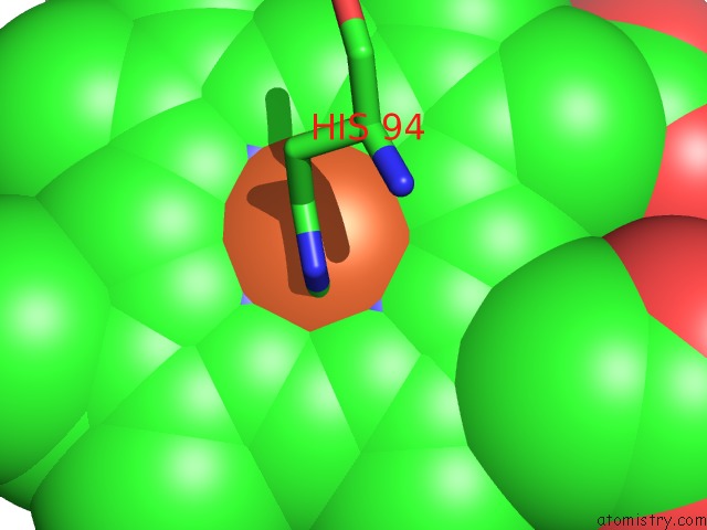
Mono view
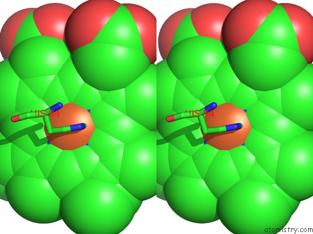
Stereo pair view
Mono view
Stereo pair view
A full contact list of Iron with other atoms in the Fe binding
site number 1 of Structure of the Partially Unliganded Met State of 400 kDa Hemoglobin: Insights Into Ligand-Induced Structural Changes of Giant Hemoglobins within 5.0Å range:
|
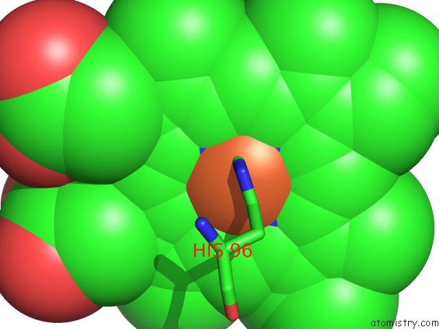
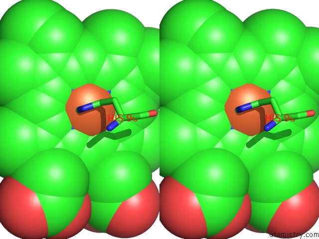
Iron binding site 2 out of 4 in 2zfo
Go back to
Iron binding site 2 out
of 4 in the Structure of the Partially Unliganded Met State of 400 kDa Hemoglobin: Insights Into Ligand-Induced Structural Changes of Giant Hemoglobins
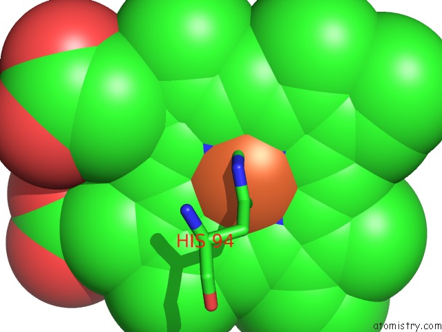
Mono view
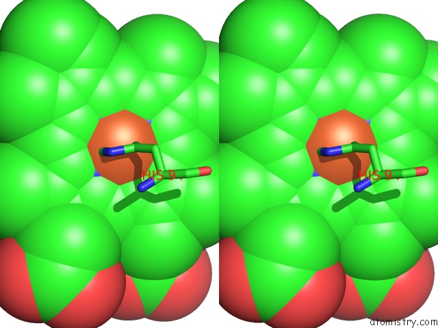
Stereo pair view
Mono view
Stereo pair view
A full contact list of Iron with other atoms in the Fe binding
site number 2 of Structure of the Partially Unliganded Met State of 400 kDa Hemoglobin: Insights Into Ligand-Induced Structural Changes of Giant Hemoglobins within 5.0Å range:
|
Iron binding site 3 out of 4 in 2zfo
Go back to
Iron binding site 3 out
of 4 in the Structure of the Partially Unliganded Met State of 400 kDa Hemoglobin: Insights Into Ligand-Induced Structural Changes of Giant Hemoglobins
Mono view
Stereo pair view
Mono view
Stereo pair view
A full contact list of Iron with other atoms in the Fe binding
site number 3 of Structure of the Partially Unliganded Met State of 400 kDa Hemoglobin: Insights Into Ligand-Induced Structural Changes of Giant Hemoglobins within 5.0Å range:
|
Iron binding site 4 out of 4 in 2zfo
Go back to
Iron binding site 4 out
of 4 in the Structure of the Partially Unliganded Met State of 400 kDa Hemoglobin: Insights Into Ligand-Induced Structural Changes of Giant Hemoglobins
Mono view
Stereo pair view
Mono view
Stereo pair view
A full contact list of Iron with other atoms in the Fe binding
site number 4 of Structure of the Partially Unliganded Met State of 400 kDa Hemoglobin: Insights Into Ligand-Induced Structural Changes of Giant Hemoglobins within 5.0Å range:
|
Reference:
N.Numoto,
T.Nakagawa,
A.Kita,
Y.Sasayama,
Y.Fukumori,
K.Miki.
Structure of the Partially Unliganded Met State of 400 kDa Hemoglobin: Insights Into Ligand-Induced Structural Changes of Giant Hemoglobins Proteins V. 73 113 2008.
ISSN: ISSN 0887-3585
PubMed: 18398907
DOI: 10.1002/PROT.22040
Page generated: Mon Aug 4 22:45:58 2025
ISSN: ISSN 0887-3585
PubMed: 18398907
DOI: 10.1002/PROT.22040
Last articles
Mn in 9LJUMn in 9LJW
Mn in 9LJS
Mn in 9LJR
Mn in 9LJT
Mn in 9LJV
Mg in 9UA2
Mg in 9R96
Mg in 9VM1
Mg in 9P01