Iron »
PDB 2z5z-2zpg »
2zi8 »
Iron in PDB 2zi8: Crystal Structure of the Hsac Extradiol Dioxygenase From M. Tuberculosis in Complex with 3,4-Dihydroxy-9,10- Seconandrost-1,3,5(10)-Triene-9,17-Dione (Dhsa)
Enzymatic activity of Crystal Structure of the Hsac Extradiol Dioxygenase From M. Tuberculosis in Complex with 3,4-Dihydroxy-9,10- Seconandrost-1,3,5(10)-Triene-9,17-Dione (Dhsa)
All present enzymatic activity of Crystal Structure of the Hsac Extradiol Dioxygenase From M. Tuberculosis in Complex with 3,4-Dihydroxy-9,10- Seconandrost-1,3,5(10)-Triene-9,17-Dione (Dhsa):
1.13.11.39;
1.13.11.39;
Protein crystallography data
The structure of Crystal Structure of the Hsac Extradiol Dioxygenase From M. Tuberculosis in Complex with 3,4-Dihydroxy-9,10- Seconandrost-1,3,5(10)-Triene-9,17-Dione (Dhsa), PDB code: 2zi8
was solved by
I.D'angelo,
K.C.Yam,
L.D.Eltis,
N.Strynadka,
with X-Ray Crystallography technique. A brief refinement statistics is given in the table below:
| Resolution Low / High (Å) | 20.00 / 2.20 |
| Space group | P 4 21 2 |
| Cell size a, b, c (Å), α, β, γ (°) | 124.318, 124.318, 106.383, 90.00, 90.00, 90.00 |
| R / Rfree (%) | 19.4 / 26.4 |
Iron Binding Sites:
The binding sites of Iron atom in the Crystal Structure of the Hsac Extradiol Dioxygenase From M. Tuberculosis in Complex with 3,4-Dihydroxy-9,10- Seconandrost-1,3,5(10)-Triene-9,17-Dione (Dhsa)
(pdb code 2zi8). This binding sites where shown within
5.0 Angstroms radius around Iron atom.
In total 2 binding sites of Iron where determined in the Crystal Structure of the Hsac Extradiol Dioxygenase From M. Tuberculosis in Complex with 3,4-Dihydroxy-9,10- Seconandrost-1,3,5(10)-Triene-9,17-Dione (Dhsa), PDB code: 2zi8:
Jump to Iron binding site number: 1; 2;
In total 2 binding sites of Iron where determined in the Crystal Structure of the Hsac Extradiol Dioxygenase From M. Tuberculosis in Complex with 3,4-Dihydroxy-9,10- Seconandrost-1,3,5(10)-Triene-9,17-Dione (Dhsa), PDB code: 2zi8:
Jump to Iron binding site number: 1; 2;
Iron binding site 1 out of 2 in 2zi8
Go back to
Iron binding site 1 out
of 2 in the Crystal Structure of the Hsac Extradiol Dioxygenase From M. Tuberculosis in Complex with 3,4-Dihydroxy-9,10- Seconandrost-1,3,5(10)-Triene-9,17-Dione (Dhsa)
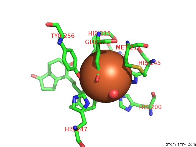
Mono view
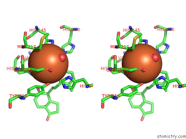
Stereo pair view
Mono view
Stereo pair view
A full contact list of Iron with other atoms in the Fe binding
site number 1 of Crystal Structure of the Hsac Extradiol Dioxygenase From M. Tuberculosis in Complex with 3,4-Dihydroxy-9,10- Seconandrost-1,3,5(10)-Triene-9,17-Dione (Dhsa) within 5.0Å range:
|
Iron binding site 2 out of 2 in 2zi8
Go back to
Iron binding site 2 out
of 2 in the Crystal Structure of the Hsac Extradiol Dioxygenase From M. Tuberculosis in Complex with 3,4-Dihydroxy-9,10- Seconandrost-1,3,5(10)-Triene-9,17-Dione (Dhsa)
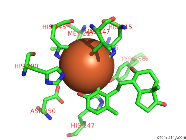
Mono view
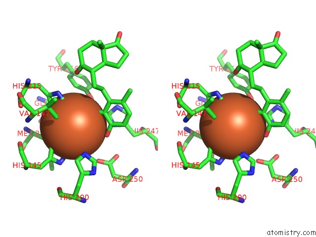
Stereo pair view
Mono view
Stereo pair view
A full contact list of Iron with other atoms in the Fe binding
site number 2 of Crystal Structure of the Hsac Extradiol Dioxygenase From M. Tuberculosis in Complex with 3,4-Dihydroxy-9,10- Seconandrost-1,3,5(10)-Triene-9,17-Dione (Dhsa) within 5.0Å range:
|
Reference:
K.C.Yam,
I.D'angelo,
R.Kalscheuer,
H.Zhu,
J.X.Wang,
V.Snieckus,
L.H.Ly,
P.J.Converse,
W.R.Jacobs,
N.Strynadka,
L.D.Eltis.
Studies of A Ring-Cleaving Dioxygenase Illuminate the Role of Cholesterol Metabolism in the Pathogenesis of Mycobacterium Tuberculosis. Plos Pathog. V. 5 E1000 2009.
ISSN: ISSN 1553-7366
PubMed: 19300498
DOI: 10.1371/JOURNAL.PPAT.1000344
Page generated: Mon Aug 4 22:46:58 2025
ISSN: ISSN 1553-7366
PubMed: 19300498
DOI: 10.1371/JOURNAL.PPAT.1000344
Last articles
Mn in 9LJUMn in 9LJW
Mn in 9LJS
Mn in 9LJR
Mn in 9LJT
Mn in 9LJV
Mg in 9UA2
Mg in 9R96
Mg in 9VM1
Mg in 9P01