Iron »
PDB 2zph-3a0b »
2zt2 »
Iron in PDB 2zt2: Carbonmonoxy Sperm Whale Myoglobin at 120 K: Laser on [600 Min]
Protein crystallography data
The structure of Carbonmonoxy Sperm Whale Myoglobin at 120 K: Laser on [600 Min], PDB code: 2zt2
was solved by
A.Tomita,
T.Sato,
K.Ichiyanagi,
S.Nozawa,
H.Ichikawa,
M.Chollet,
F.Kawai,
S.-Y.Park,
S.Koshihara,
S.Adachi,
with X-Ray Crystallography technique. A brief refinement statistics is given in the table below:
| Resolution Low / High (Å) | 20.01 / 1.21 |
| Space group | P 1 21 1 |
| Cell size a, b, c (Å), α, β, γ (°) | 34.453, 30.718, 63.907, 90.00, 105.59, 90.00 |
| R / Rfree (%) | 14.4 / 18.6 |
Iron Binding Sites:
The binding sites of Iron atom in the Carbonmonoxy Sperm Whale Myoglobin at 120 K: Laser on [600 Min]
(pdb code 2zt2). This binding sites where shown within
5.0 Angstroms radius around Iron atom.
In total only one binding site of Iron was determined in the Carbonmonoxy Sperm Whale Myoglobin at 120 K: Laser on [600 Min], PDB code: 2zt2:
In total only one binding site of Iron was determined in the Carbonmonoxy Sperm Whale Myoglobin at 120 K: Laser on [600 Min], PDB code: 2zt2:
Iron binding site 1 out of 1 in 2zt2
Go back to
Iron binding site 1 out
of 1 in the Carbonmonoxy Sperm Whale Myoglobin at 120 K: Laser on [600 Min]
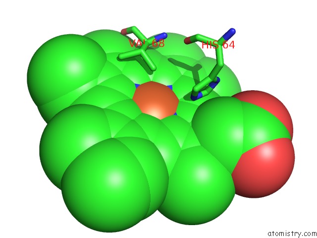
Mono view
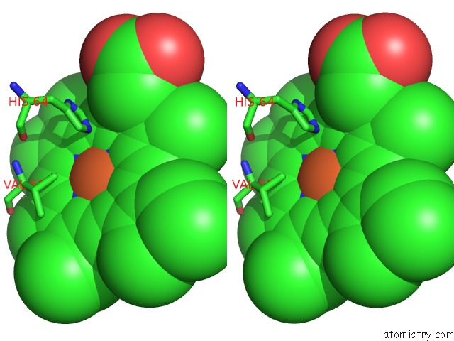
Stereo pair view
Mono view
Stereo pair view
A full contact list of Iron with other atoms in the Fe binding
site number 1 of Carbonmonoxy Sperm Whale Myoglobin at 120 K: Laser on [600 Min] within 5.0Å range:
|
Reference:
A.Tomita,
T.Sato,
K.Ichiyanagi,
S.Nozawa,
H.Ichikawa,
M.Chollet,
F.Kawai,
S.-Y.Park,
T.Tsuduki,
T.Yamato,
S.Koshihara,
S.Adachi.
Visualizing Breathing Motion of Internal Cavities in Concert with Ligand Migration in Myoglobin Proc.Natl.Acad.Sci.Usa V. 106 2612 2009.
ISSN: ISSN 0027-8424
PubMed: 19204297
DOI: 10.1073/PNAS.0807774106
Page generated: Mon Aug 4 22:56:25 2025
ISSN: ISSN 0027-8424
PubMed: 19204297
DOI: 10.1073/PNAS.0807774106
Last articles
Mn in 9LJUMn in 9LJW
Mn in 9LJS
Mn in 9LJR
Mn in 9LJT
Mn in 9LJV
Mg in 9UA2
Mg in 9R96
Mg in 9VM1
Mg in 9P01